


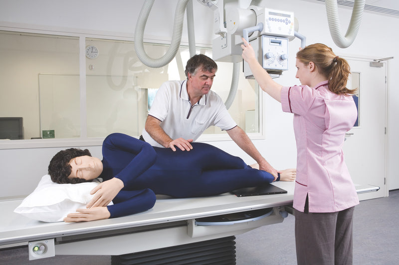
El maniquí para posicionamiento radiográfico con rayos X es un simulador de entrenamiento de tamaño real diseñado para enseñar el posicionamiento radiográfico, la manipulación de pacientes y los procedimientos de obtención de imágenes sin exponer a pacientes ni estudiantes a la radiación. Utilizado ampliamente por los programas de radiografía, este simulador permite a los estudiantes practicar técnicas de posicionamiento correctas en un entorno seguro y controlado.
El maniquí contiene un esqueleto completamente articulado fabricado con plástico duradero y sin componentes metálicos, lo que permite utilizarlo durante estudios de rayos X y tomografías computarizadas. Las estructuras internas radiolúcidas y la anatomía superficial realista facilitan la identificación de los puntos de referencia anatómicos durante la capacitación en posicionamiento y obtención de imágenes.
Características principales
• Maniquí de tamaño real para posicionamiento radiográfico
• Esqueleto completamente flexible y articulado
• Sin piezas metálicas para un uso seguro con rayos X y tomografía computarizada
• Órganos internos y estructuras anatómicas radiolúcidos
• Flexibilidad articular realista para practicar el posicionamiento
• Construcción duradera para usos repetidos en capacitación
• Ligero para facilitar su manipulación
Aplicaciones de capacitación
• Técnicas de posicionamiento radiográfico
• Capacitación en manipulación de pacientes
• Posicionamiento en equipos de rayos X
• Práctica de posicionamiento para tomografía computarizada
• Instrucción en laboratorios de obtención de imágenes
• Programas educativos de radiografía
Detalles adicionales
• Se suministra con imágenes individuales de rayos X del maniquí
• Adecuado para practicar el posicionamiento de todo el cuerpo
• Diseñado para usos repetidos en el aula
Garantía de 2 años.
Peso de envío: 38 lb
Dimensiones (pulgadas): 68.5 x 20.5 x 14
Key Features
• Life-size radiographic positioning manikin
• Fully flexible articulated skeleton
• No metal parts for safe use with X-ray and CT
• Radiolucent internal organs and anatomical structures
• Realistic joint flexibility for positioning practice
• Durable construction for repeated training use
• Lightweight for easy handling
Downloads
Individual X-Rays
Individual X-Rays for each Doll
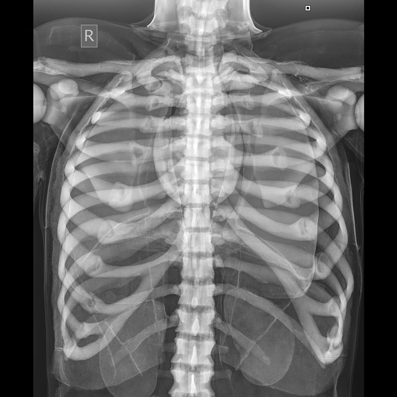
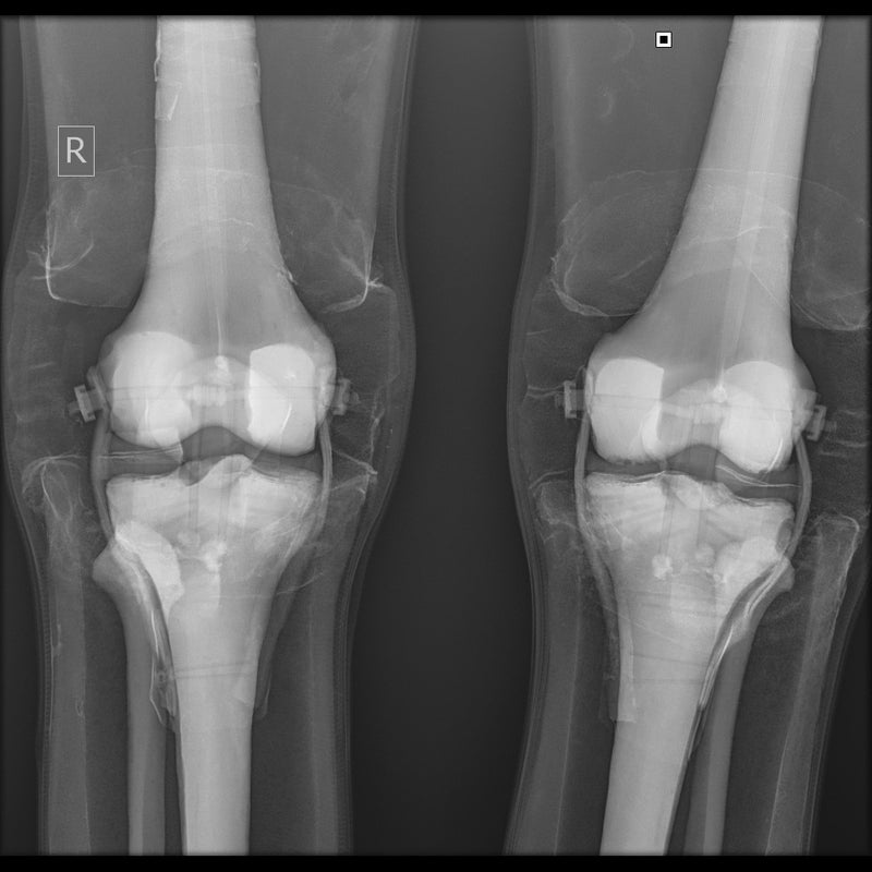
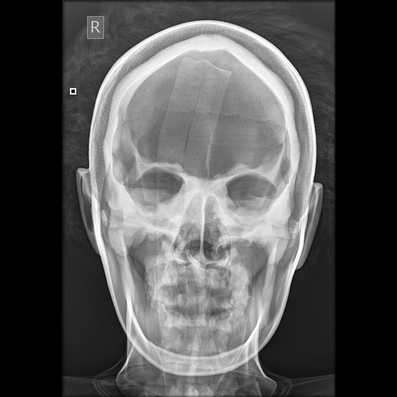
- Each Doll is individually X-rayed
- Customers can be reassured that there are no metal parts in the Doll by referring to the set of digital X-
- Rays which are supplied with each Doll
- Images shown were taken using medium frequency generator; no grid; fine focus; 100cm S.I.D. Fuji CR system
Positioning and CT Scanning
Positioning
- The Doll is best used in the recumbent positions as, although it is possible to use an upright bucky with supports, this makes the use of positioning blocks and immobilization devices more difficult
- The Doll will lie naturally in the neutral position, prone or supine, without support
- A wide selection of positioning aids must be at hand, e.g. foam blocks, wedges, sand bags and compression band. These are essential to maintain any position other than neutral as the skin of the Doll has some resistance to overcome. This is, of course, an advantage in the teaching as it prevents the student cutting corners by omitting the use of these positioning aids
- The Doll can be positioned for all standard projections with no more difficulty than a difficult patient might present.
CT Scanning
- The X-Ray Positioning Doll was positioned and imaged for a whole body CT (Computed Tomography) investigation
- Good images in terms of body outline and fundamental structure can be obtained
- Certain areas of the skeleton structure e.g. thoracic vertebrae and the head of the femur gave similar values to the typical patient
- As the doll is very different from the human body in terms of attenuation, it cannot be used for training in, or practising exposure control
El maniquí para posicionamiento radiográfico con rayos X es un simulador de entrenamiento de tamaño real diseñado para enseñar el posicionamiento radiográfico, la manipulación de pacientes y los procedimientos de obtención de imágenes sin exponer a pacientes ni estudiantes a la radiación. Utilizado ampliamente por los programas de radiografía, este simulador permite a los estudiantes practicar técnicas de posicionamiento correctas en un entorno seguro y controlado.
El maniquí contiene un esqueleto completamente articulado fabricado con plástico duradero y sin componentes metálicos, lo que permite utilizarlo durante estudios de rayos X y tomografías computarizadas. Las estructuras internas radiolúcidas y la anatomía superficial realista facilitan la identificación de los puntos de referencia anatómicos durante la capacitación en posicionamiento y obtención de imágenes.
Características principales
• Maniquí de tamaño real para posicionamiento radiográfico
• Esqueleto completamente flexible y articulado
• Sin piezas metálicas para un uso seguro con rayos X y tomografía computarizada
• Órganos internos y estructuras anatómicas radiolúcidos
• Flexibilidad articular realista para practicar el posicionamiento
• Construcción duradera para usos repetidos en capacitación
• Ligero para facilitar su manipulación
Aplicaciones de capacitación
• Técnicas de posicionamiento radiográfico
• Capacitación en manipulación de pacientes
• Posicionamiento en equipos de rayos X
• Práctica de posicionamiento para tomografía computarizada
• Instrucción en laboratorios de obtención de imágenes
• Programas educativos de radiografía
Detalles adicionales
• Se suministra con imágenes individuales de rayos X del maniquí
• Adecuado para practicar el posicionamiento de todo el cuerpo
• Diseñado para usos repetidos en el aula
Garantía de 2 años.
Peso de envío: 38 lb
Dimensiones (pulgadas): 68.5 x 20.5 x 14
Key Features
• Life-size radiographic positioning manikin
• Fully flexible articulated skeleton
• No metal parts for safe use with X-ray and CT
• Radiolucent internal organs and anatomical structures
• Realistic joint flexibility for positioning practice
• Durable construction for repeated training use
• Lightweight for easy handling
Downloads
Individual X-Rays
Individual X-Rays for each Doll
- Each Doll is individually X-rayed
- Customers can be reassured that there are no metal parts in the Doll by referring to the set of digital X-
- Rays which are supplied with each Doll
- Images shown were taken using medium frequency generator; no grid; fine focus; 100cm S.I.D. Fuji CR system
Positioning and CT Scanning
Positioning
- The Doll is best used in the recumbent positions as, although it is possible to use an upright bucky with supports, this makes the use of positioning blocks and immobilization devices more difficult
- The Doll will lie naturally in the neutral position, prone or supine, without support
- A wide selection of positioning aids must be at hand, e.g. foam blocks, wedges, sand bags and compression band. These are essential to maintain any position other than neutral as the skin of the Doll has some resistance to overcome. This is, of course, an advantage in the teaching as it prevents the student cutting corners by omitting the use of these positioning aids
- The Doll can be positioned for all standard projections with no more difficulty than a difficult patient might present.
CT Scanning
- The X-Ray Positioning Doll was positioned and imaged for a whole body CT (Computed Tomography) investigation
- Good images in terms of body outline and fundamental structure can be obtained
- Certain areas of the skeleton structure e.g. thoracic vertebrae and the head of the femur gave similar values to the typical patient
- As the doll is very different from the human body in terms of attenuation, it cannot be used for training in, or practising exposure control