


วิลไลในลำไส้ ขยาย 100 เท่า
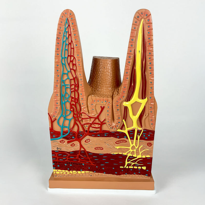
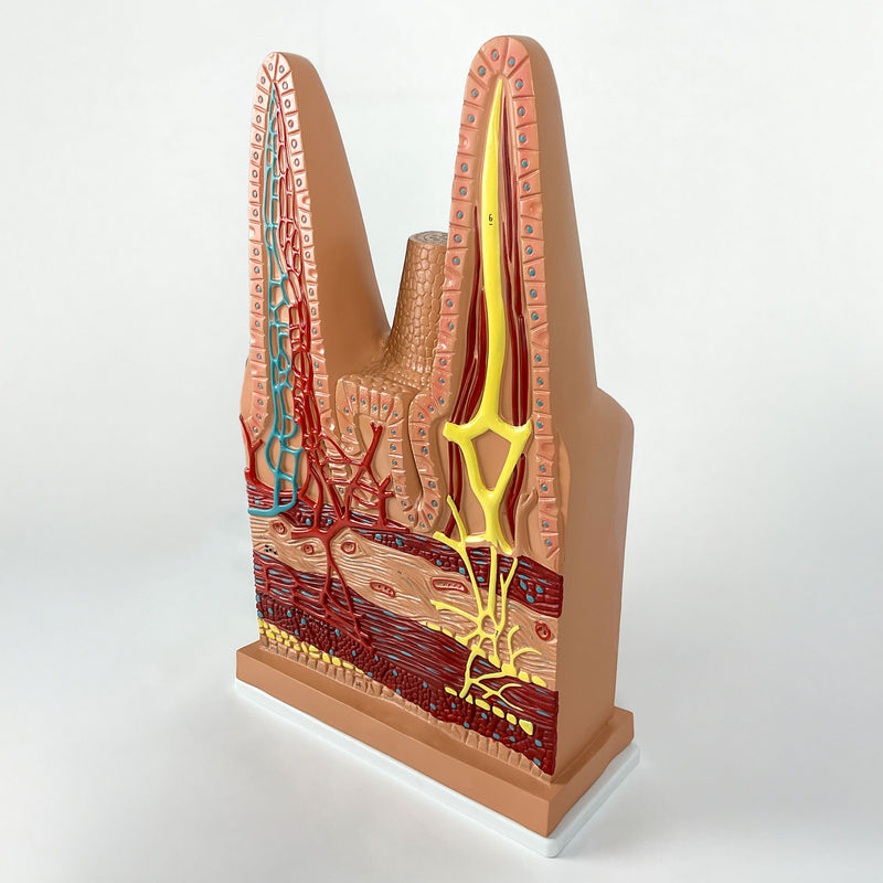
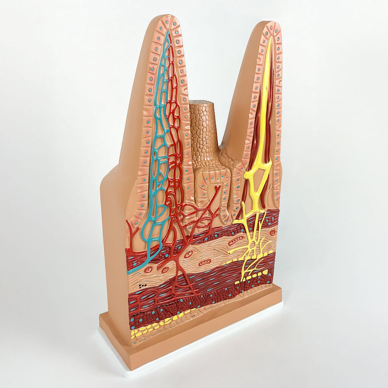
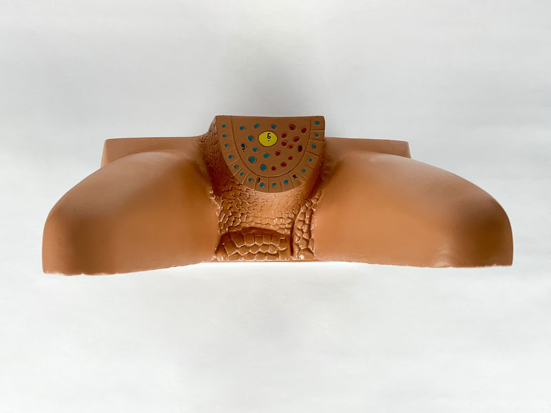
แบบจำลองทางกายวิภาคของวิลไลในลำไส้ที่ขยายให้ใหญ่ขึ้น 100 เท่า แสดงรายละเอียดของวิลไล 3 อัน ได้แก่ วิลไลที่สมบูรณ์ 1 อัน และวิลไลที่ผ่าตัดตามขวาง 2 อัน เพื่อแสดงหลอดเลือด หลอดน้ำเหลือง เส้นเลือดฝอย และเซลล์เยื่อบุผิว แบบจำลองทาสีด้วยมือและมีหมายเลขกำกับ ช่วยให้ศึกษาโครงสร้างต่าง ๆ ได้สะดวก
สัมผัสประสบการณ์ความเป็นจริงเสริมผ่านบัตรข้อมูลที่ให้มา พร้อมสำรวจแบบจำลอง 3 มิติแบบโต้ตอบ เพื่อการเรียนรู้กายวิภาคและสรีรวิทยาของลำไส้ที่ดียิ่งขึ้น เหมาะสำหรับนักศึกษาและบุคลากรทางการแพทย์
การเรียนรู้ผ่าน 3 มิติแบบโต้ตอบและความเป็นจริงเสริม
ใช้แหล่งข้อมูล 3 มิติแบบโต้ตอบและความเป็นจริงเสริมที่มาพร้อมกัน หากมี เพื่อศึกษาตรวจสอบและเปรียบเทียบโครงสร้างทางกายวิภาคได้อย่างละเอียดมากยิ่งขึ้น
Intestinal Villi, 100 Times Enlarged Key Features
- Detailed and enlarged representation of the structures of the intestinal villus.
- Numbered and hand-painted in didactic colors.
- High-precision natural molding.
- Manufactured from stable synthetic material and resin approved in toxicological tests.
Main Structures
- 1 - Intestinal villus
- 2 - Intestinal gland
- 3 - Intestinal epithelium
- 4 - Goblet cell
- 5 - Smooth muscle
- 6 - Centual larteal
- 7 - Lymphatic vasseis
- 8 - Small antery
- 9 - Small vein
- 10 - Muscularis mucosae
Product Specifications
- Scale: 100x
- Material: Synthetic resin
- Type: Anatomical Model