


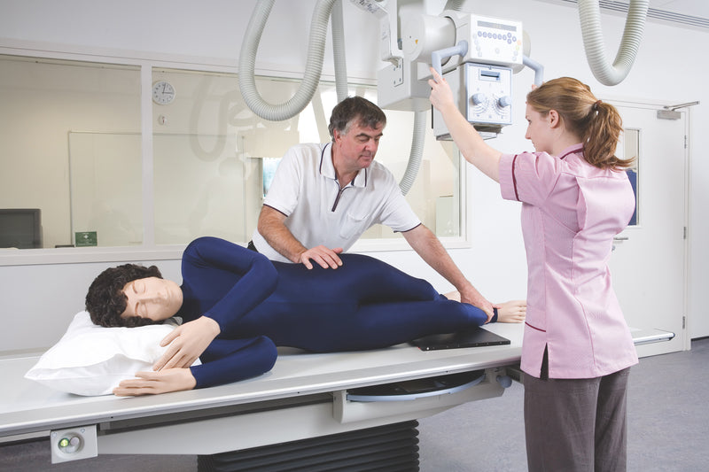
O Simulador Anatômico de Posicionamento Radiográfico para Raios X é um simulador de treinamento em tamanho real desenvolvido para o ensino de posicionamento radiográfico, manejo de pacientes e procedimentos de imagem sem expor pacientes ou estudantes à radiação. Amplamente utilizado por programas de radiografia, este simulador permite que os alunos pratiquem técnicas corretas de posicionamento em um ambiente seguro e controlado.
O simulador contém um esqueleto totalmente articulado, feito de plástico durável e sem componentes metálicos, permitindo seu uso durante exames de raios X e tomografia computadorizada. As estruturas internas radiotransparentes e a anatomia superficial realista facilitam a identificação de referências anatômicas durante o treinamento de posicionamento e aquisição de imagens.
Principais características
• Simulador de posicionamento radiográfico em tamanho real
• Esqueleto totalmente flexível e articulado
• Sem peças metálicas, para uso seguro com raios X e tomografia computadorizada
• Órgãos internos e estruturas anatômicas radiotransparentes
• Flexibilidade articular realista para a prática de posicionamento
• Construção durável para uso repetido em treinamentos
• Leve, para facilitar o manuseio
Aplicações de treinamento
• Técnicas de posicionamento radiográfico
• Treinamento de manejo de pacientes
• Posicionamento em equipamentos de raios X
• Prática de posicionamento para tomografia computadorizada
• Ensino em laboratórios de diagnóstico por imagem
• Programas de formação em radiografia
Detalhes adicionais
• Fornecido com imagens individuais de raios X do simulador
• Adequado para a prática de posicionamento de corpo inteiro
• Desenvolvido para uso repetido em sala de aula
Garantia de 2 anos.
Peso para envio: 38 lb
Dimensões (polegadas): 68.5 x 20.5 x 14
Key Features
• Life-size radiographic positioning manikin
• Fully flexible articulated skeleton
• No metal parts for safe use with X-ray and CT
• Radiolucent internal organs and anatomical structures
• Realistic joint flexibility for positioning practice
• Durable construction for repeated training use
• Lightweight for easy handling
Downloads
Individual X-Rays
Individual X-Rays for each Doll
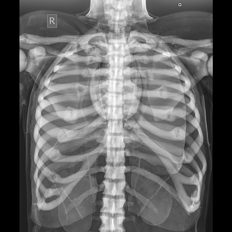
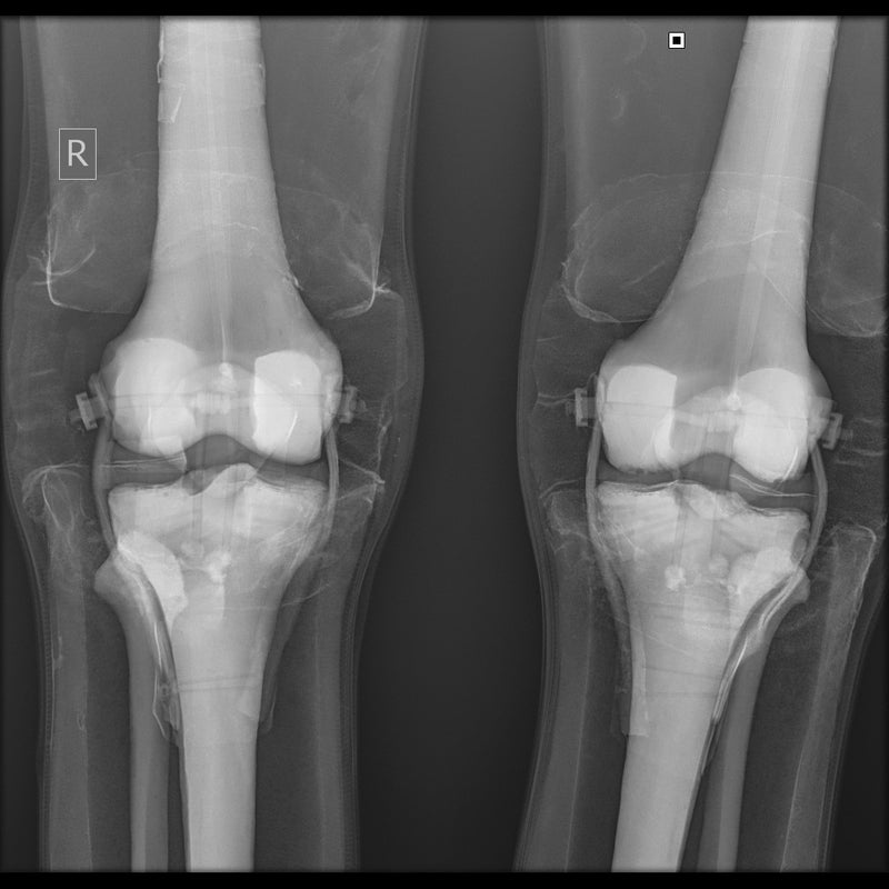
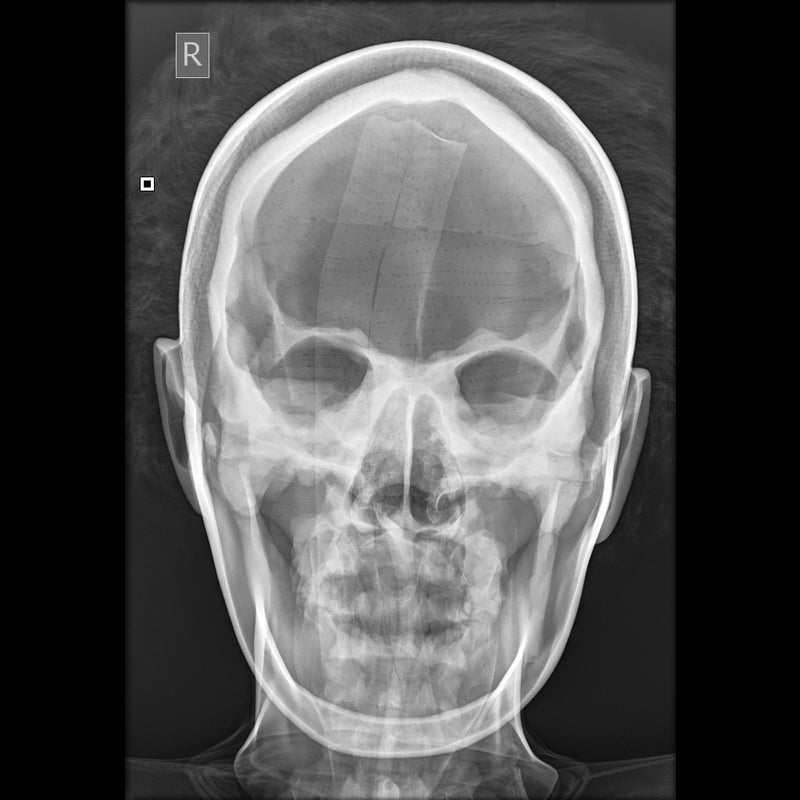
- Each Doll is individually X-rayed
- Customers can be reassured that there are no metal parts in the Doll by referring to the set of digital X-
- Rays which are supplied with each Doll
- Images shown were taken using medium frequency generator; no grid; fine focus; 100cm S.I.D. Fuji CR system
Positioning and CT Scanning
Positioning
- The Doll is best used in the recumbent positions as, although it is possible to use an upright bucky with supports, this makes the use of positioning blocks and immobilization devices more difficult
- The Doll will lie naturally in the neutral position, prone or supine, without support
- A wide selection of positioning aids must be at hand, e.g. foam blocks, wedges, sand bags and compression band. These are essential to maintain any position other than neutral as the skin of the Doll has some resistance to overcome. This is, of course, an advantage in the teaching as it prevents the student cutting corners by omitting the use of these positioning aids
- The Doll can be positioned for all standard projections with no more difficulty than a difficult patient might present.
CT Scanning
- The X-Ray Positioning Doll was positioned and imaged for a whole body CT (Computed Tomography) investigation
- Good images in terms of body outline and fundamental structure can be obtained
- Certain areas of the skeleton structure e.g. thoracic vertebrae and the head of the femur gave similar values to the typical patient
- As the doll is very different from the human body in terms of attenuation, it cannot be used for training in, or practising exposure control
O Simulador Anatômico de Posicionamento Radiográfico para Raios X é um simulador de treinamento em tamanho real desenvolvido para o ensino de posicionamento radiográfico, manejo de pacientes e procedimentos de imagem sem expor pacientes ou estudantes à radiação. Amplamente utilizado por programas de radiografia, este simulador permite que os alunos pratiquem técnicas corretas de posicionamento em um ambiente seguro e controlado.
O simulador contém um esqueleto totalmente articulado, feito de plástico durável e sem componentes metálicos, permitindo seu uso durante exames de raios X e tomografia computadorizada. As estruturas internas radiotransparentes e a anatomia superficial realista facilitam a identificação de referências anatômicas durante o treinamento de posicionamento e aquisição de imagens.
Principais características
• Simulador de posicionamento radiográfico em tamanho real
• Esqueleto totalmente flexível e articulado
• Sem peças metálicas, para uso seguro com raios X e tomografia computadorizada
• Órgãos internos e estruturas anatômicas radiotransparentes
• Flexibilidade articular realista para a prática de posicionamento
• Construção durável para uso repetido em treinamentos
• Leve, para facilitar o manuseio
Aplicações de treinamento
• Técnicas de posicionamento radiográfico
• Treinamento de manejo de pacientes
• Posicionamento em equipamentos de raios X
• Prática de posicionamento para tomografia computadorizada
• Ensino em laboratórios de diagnóstico por imagem
• Programas de formação em radiografia
Detalhes adicionais
• Fornecido com imagens individuais de raios X do simulador
• Adequado para a prática de posicionamento de corpo inteiro
• Desenvolvido para uso repetido em sala de aula
Garantia de 2 anos.
Peso para envio: 38 lb
Dimensões (polegadas): 68.5 x 20.5 x 14
Key Features
• Life-size radiographic positioning manikin
• Fully flexible articulated skeleton
• No metal parts for safe use with X-ray and CT
• Radiolucent internal organs and anatomical structures
• Realistic joint flexibility for positioning practice
• Durable construction for repeated training use
• Lightweight for easy handling
Downloads
Individual X-Rays
Individual X-Rays for each Doll
- Each Doll is individually X-rayed
- Customers can be reassured that there are no metal parts in the Doll by referring to the set of digital X-
- Rays which are supplied with each Doll
- Images shown were taken using medium frequency generator; no grid; fine focus; 100cm S.I.D. Fuji CR system
Positioning and CT Scanning
Positioning
- The Doll is best used in the recumbent positions as, although it is possible to use an upright bucky with supports, this makes the use of positioning blocks and immobilization devices more difficult
- The Doll will lie naturally in the neutral position, prone or supine, without support
- A wide selection of positioning aids must be at hand, e.g. foam blocks, wedges, sand bags and compression band. These are essential to maintain any position other than neutral as the skin of the Doll has some resistance to overcome. This is, of course, an advantage in the teaching as it prevents the student cutting corners by omitting the use of these positioning aids
- The Doll can be positioned for all standard projections with no more difficulty than a difficult patient might present.
CT Scanning
- The X-Ray Positioning Doll was positioned and imaged for a whole body CT (Computed Tomography) investigation
- Good images in terms of body outline and fundamental structure can be obtained
- Certain areas of the skeleton structure e.g. thoracic vertebrae and the head of the femur gave similar values to the typical patient
- As the doll is very different from the human body in terms of attenuation, it cannot be used for training in, or practising exposure control