


Fertilization and Embryonic Development
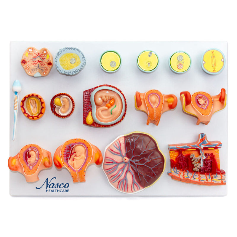
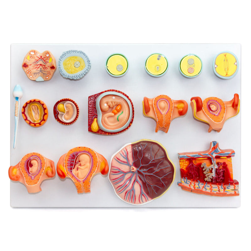
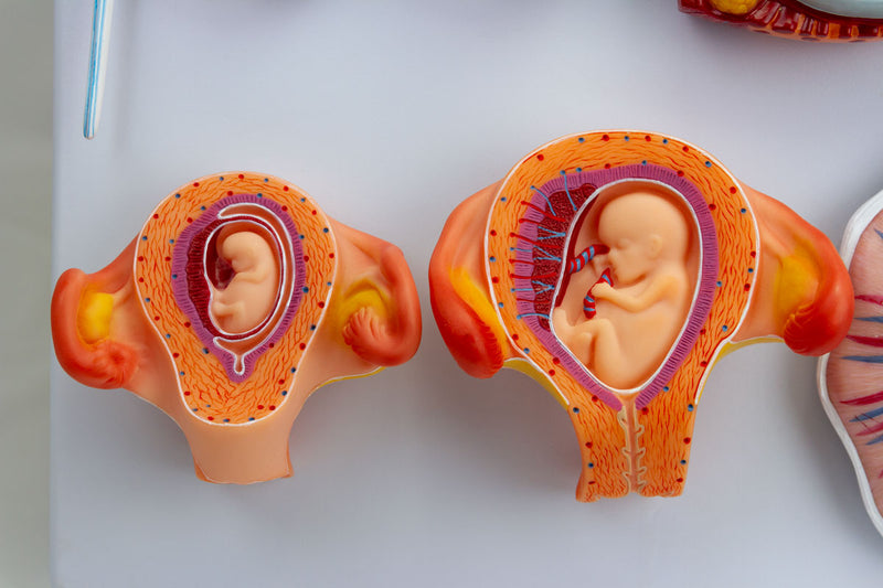
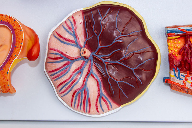
This detailed anatomical model presents 16 stages of human fertilization and embryonic development, from the union of gametes to the fetus at three months, including the placenta. Made of high-quality synthetic resin, with hand-painting and precise numbering, the model is mounted on a polymer base and includes an information card with augmented reality (AR).
Interactive 3D visualization via AR complements learning, allowing detailed analysis of structures such as pronuclei, zygote, blastoderm, and embryo at different stages. Ideal for education and research in medical and biological fields.
Interactive 3D & Augmented Reality Learning
Use the accompanying interactive 3D and augmented reality resources, where provided, to examine and compare anatomical structures in greater detail.
Fertilization and Embryonic Development Key Features
- Detailed representation of each stage of embryonic development.
- Allows the study of the structures that make up the placenta.
- High-precision natural molding.
- Made of stable and resistant synthetic material.
- Original, numbered, and hand-painted replicas.
- Resin approved in toxicological tests.
Main Structures
- Mitosis
- Female pronucleus close to male pronucleus
- Female pronucleus and male pronucleus
- Amphimixis or zygote
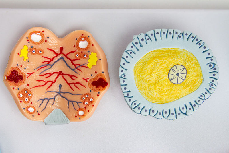
- Ovum
- Ovary
- Spermatozoon
- Blastoderm
- Embryo at week 2-3
- Embryo at week 5
Product Specifications
- Material: Synthetic resin.
- Scale: Unspecified.
- Manual numbering and painting.
- Information card included.
Fertilization and Embryonic Development
This detailed anatomical model presents 16 stages of human fertilization and embryonic development, from the union of gametes to the fetus at three months, including the placenta. Made of high-quality synthetic resin, with hand-painting and precise numbering, the model is mounted on a polymer base and includes an information card with augmented reality (AR).
Interactive 3D visualization via AR complements learning, allowing detailed analysis of structures such as pronuclei, zygote, blastoderm, and embryo at different stages. Ideal for education and research in medical and biological fields.
Interactive 3D & Augmented Reality Learning
Use the accompanying interactive 3D and augmented reality resources, where provided, to examine and compare anatomical structures in greater detail.
Fertilization and Embryonic Development Key Features
- Detailed representation of each stage of embryonic development.
- Allows the study of the structures that make up the placenta.
- High-precision natural molding.
- Made of stable and resistant synthetic material.
- Original, numbered, and hand-painted replicas.
- Resin approved in toxicological tests.
Main Structures
- Mitosis
- Female pronucleus close to male pronucleus
- Female pronucleus and male pronucleus
- Amphimixis or zygote
- Ovum
- Ovary
- Spermatozoon
- Blastoderm
- Embryo at week 2-3
- Embryo at week 5
Product Specifications
- Material: Synthetic resin.
- Scale: Unspecified.
- Manual numbering and painting.
- Information card included.