


Embryonic Development, 13 Stages
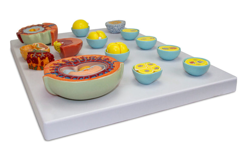
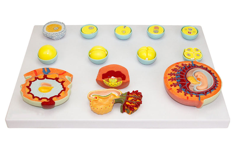
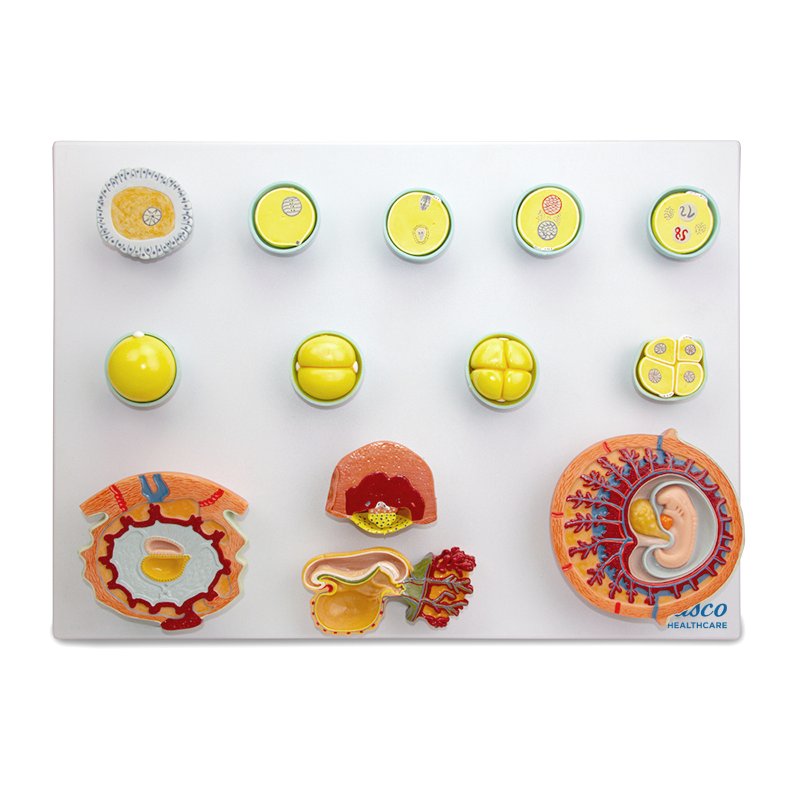
This detailed anatomical model shows the 13 initial stages of human embryonic development, from fertilization to the fourth week, with numbered and hand-painted pieces. Ideal for students and professionals in biology, medicine, and related fields, it offers an accurate and magnified representation of the main structures, in a resistant and non-toxic material.
It includes an information card with augmented reality (AR), allowing for interactive 3D visualization for enhanced learning and comparative analysis of the structures.
Interactive 3D & Augmented Reality Learning
Use the accompanying interactive 3D and augmented reality resources, where provided, to examine and compare anatomical structures in greater detail.
Embryonic Development, 13 Stages Key Features
- Precise and detailed representation of the 13 stages of embryonic development.
- Numbered and hand-painted parts for easy identification of structures.
- Manufactured from stable and resistant synthetic material, with resin approved in
- High anatomical fidelity natural molding.
- Includes an information card with related structures.
- Original replicas, enlarged from natural size.
- Model mounted on a polymer base.
Main Structures
- Ovum
- Sperm entry
- Formation of polocyte II
- Approximation of pronuclei
- Union of pronuclei (Karyogamy)
- 16-cell stage - Morula (72 hours)
- 4-cell stage - cleavage (48 hours)
- 2-cell stage - cleavage (36 hours)
- Unicellular stage - cleavage (zygote)
- Human embryo at 15 days
Product Specifications
- Material: Synthetic resin.
- Scale: Enlarged from natural size.
- Numbering: Manual.
- Painting: Manual.
Embryonic Development, 13 Stages
This detailed anatomical model shows the 13 initial stages of human embryonic development, from fertilization to the fourth week, with numbered and hand-painted pieces. Ideal for students and professionals in biology, medicine, and related fields, it offers an accurate and magnified representation of the main structures, in a resistant and non-toxic material.
It includes an information card with augmented reality (AR), allowing for interactive 3D visualization for enhanced learning and comparative analysis of the structures.
Interactive 3D & Augmented Reality Learning
Use the accompanying interactive 3D and augmented reality resources, where provided, to examine and compare anatomical structures in greater detail.
Embryonic Development, 13 Stages Key Features
- Precise and detailed representation of the 13 stages of embryonic development.
- Numbered and hand-painted parts for easy identification of structures.
- Manufactured from stable and resistant synthetic material, with resin approved in
- High anatomical fidelity natural molding.
- Includes an information card with related structures.
- Original replicas, enlarged from natural size.
- Model mounted on a polymer base.
Main Structures
- Ovum
- Sperm entry
- Formation of polocyte II
- Approximation of pronuclei
- Union of pronuclei (Karyogamy)
- 16-cell stage - Morula (72 hours)
- 4-cell stage - cleavage (48 hours)
- 2-cell stage - cleavage (36 hours)
- Unicellular stage - cleavage (zygote)
- Human embryo at 15 days
Product Specifications
- Material: Synthetic resin.
- Scale: Enlarged from natural size.
- Numbering: Manual.
- Painting: Manual.