


Human Auditory Bones, Tympanic and Coclea Membrane, 3 Parts
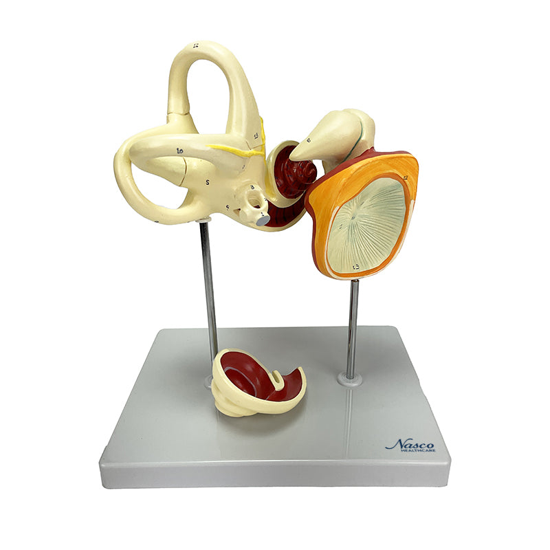
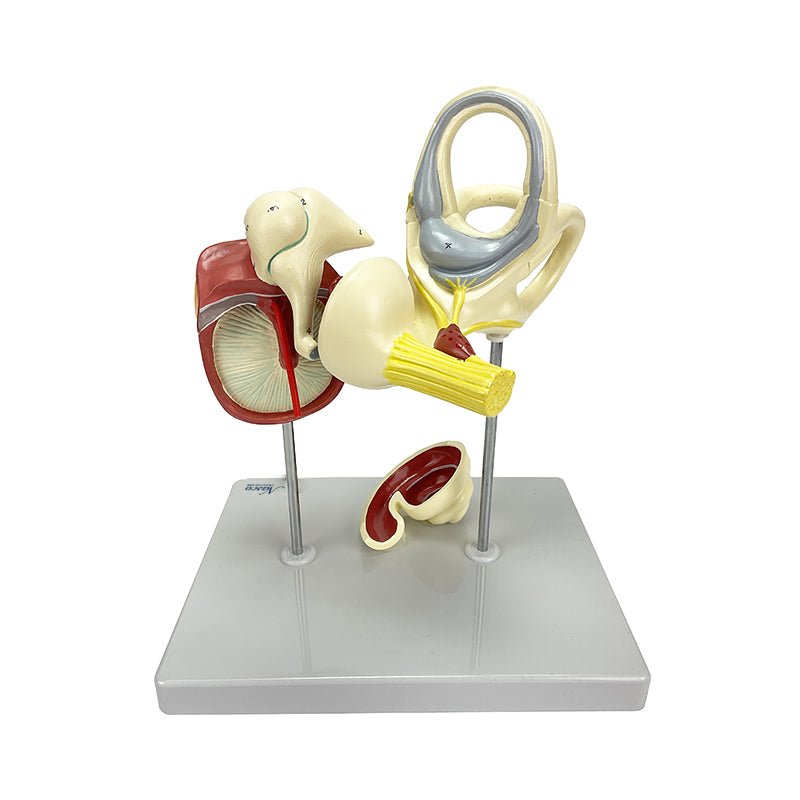
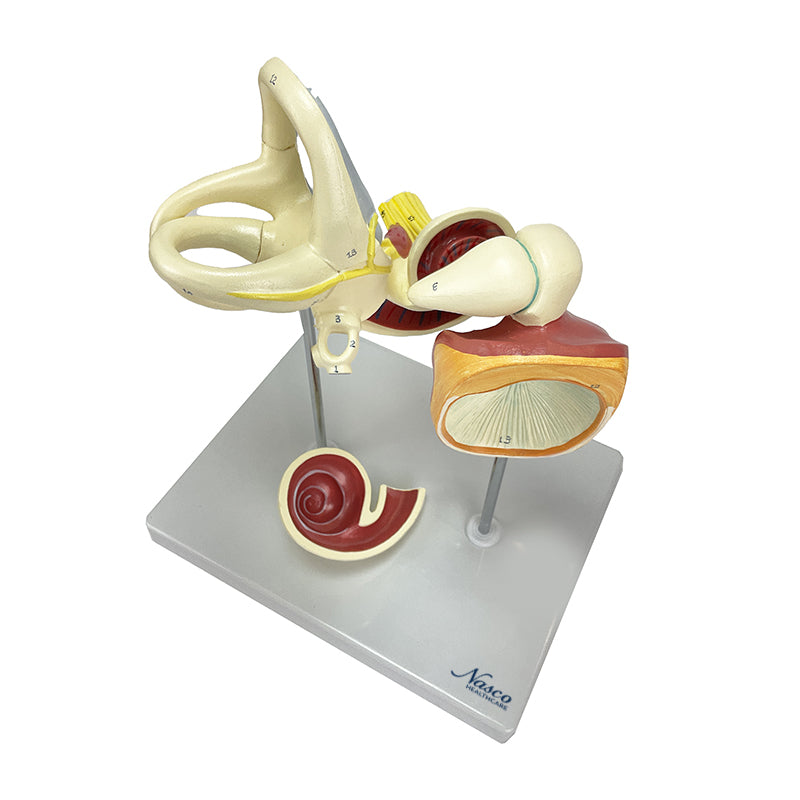
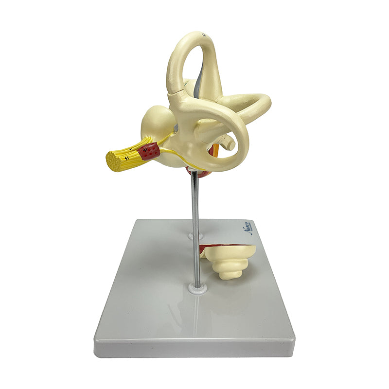
This anatomical model of the inner ear, magnified 18 times, reveals in detail the tympanic membrane, ossicles, cochlea, and vestibulocochlear nerve. Divided into three parts, it allows detailed visualization of the bony and membranous labyrinth, including the open superior semicircular canal.
Hand-painted and numbered, it includes a base and an information card with augmented reality (AR), offering an interactive learning experience for students and medical professionals. The precision of the synthetic resin molding, approved in toxicological tests, guarantees an efficient and detailed study of the anatomy of hearing and balance.
Interactive 3D & Augmented Reality Learning
Use the accompanying interactive 3D and augmented reality resources, where provided, to examine and compare anatomical structures in greater detail.
Human Auditory Bones, Tympanic and Coclea Membrane, 3 Parts Key Features
- Detailed and enlarged representation of the inner ear;
- Hand-painted in didactic colors;
- Numbered to facilitate the identification of structures;
Main Structures
- head of stapes
- crura of stapes
- base of stapes
- alveus communis
- cochlear apex
- superior bony ampullae
- cochlear nerve
- vestibular ganglion
- vestibular nerve
- posterior semicircular duct
Product Specifications
- Scale: Enlarged approximately 18 times.
- Material: Synthetic resin.
- Molding: Precision natural molding.
Human Auditory Bones, Tympanic and Coclea Membrane, 3 Parts
This anatomical model of the inner ear, magnified 18 times, reveals in detail the tympanic membrane, ossicles, cochlea, and vestibulocochlear nerve. Divided into three parts, it allows detailed visualization of the bony and membranous labyrinth, including the open superior semicircular canal.
Hand-painted and numbered, it includes a base and an information card with augmented reality (AR), offering an interactive learning experience for students and medical professionals. The precision of the synthetic resin molding, approved in toxicological tests, guarantees an efficient and detailed study of the anatomy of hearing and balance.
Interactive 3D & Augmented Reality Learning
Use the accompanying interactive 3D and augmented reality resources, where provided, to examine and compare anatomical structures in greater detail.
Human Auditory Bones, Tympanic and Coclea Membrane, 3 Parts Key Features
- Detailed and enlarged representation of the inner ear;
- Hand-painted in didactic colors;
- Numbered to facilitate the identification of structures;
Main Structures
- head of stapes
- crura of stapes
- base of stapes
- alveus communis
- cochlear apex
- superior bony ampullae
- cochlear nerve
- vestibular ganglion
- vestibular nerve
- posterior semicircular duct
Product Specifications
- Scale: Enlarged approximately 18 times.
- Material: Synthetic resin.
- Molding: Precision natural molding.