


Embryonale ontwikkeling, 13 stadia
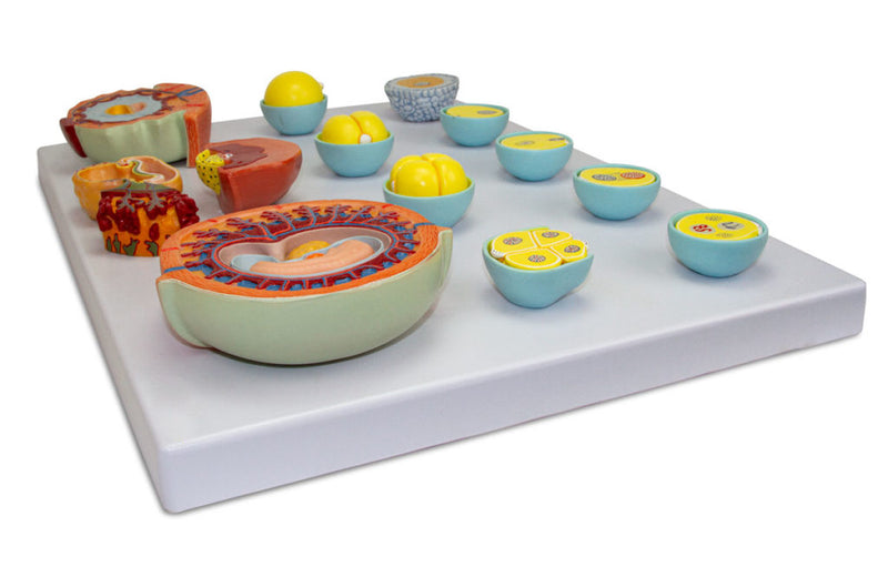
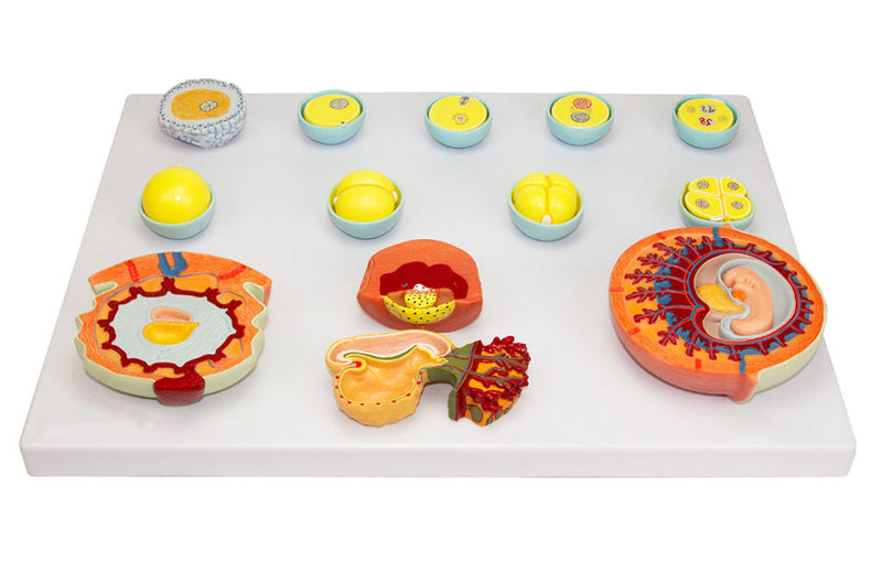
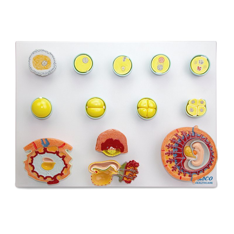
Dit gedetailleerde anatomische model toont de 13 eerste stadia van de menselijke embryonale ontwikkeling, van bevruchting tot de vierde week, met genummerde en met de hand beschilderde onderdelen. Het is ideaal voor studenten en professionals in de biologie, geneeskunde en aanverwante vakgebieden en biedt een nauwkeurige, uitvergrote weergave van de belangrijkste structuren, vervaardigd uit bestendig en niet-giftig materiaal.
Het bevat een informatiekaart met augmented reality (AR), waarmee interactieve 3D-visualisatie mogelijk is voor beter leren en een vergelijkende analyse van de structuren.
Interactief 3D- en augmented-realityleren
Gebruik de bijbehorende interactieve 3D- en augmented-realitybronnen, indien beschikbaar, om anatomische structuren in meer detail te onderzoeken en te vergelijken.
Embryonic Development, 13 Stages Key Features
- Precise and detailed representation of the 13 stages of embryonic development.
- Numbered and hand-painted parts for easy identification of structures.
- Manufactured from stable and resistant synthetic material, with resin approved in
- High anatomical fidelity natural molding.
- Includes an information card with related structures.
- Original replicas, enlarged from natural size.
- Model mounted on a polymer base.
Main Structures
- Ovum
- Sperm entry
- Formation of polocyte II
- Approximation of pronuclei
- Union of pronuclei (Karyogamy)
- 16-cell stage - Morula (72 hours)
- 4-cell stage - cleavage (48 hours)
- 2-cell stage - cleavage (36 hours)
- Unicellular stage - cleavage (zygote)
- Human embryo at 15 days
Product Specifications
- Material: Synthetic resin.
- Scale: Enlarged from natural size.
- Numbering: Manual.
- Painting: Manual.
Embryonale ontwikkeling, 13 stadia
Dit gedetailleerde anatomische model toont de 13 eerste stadia van de menselijke embryonale ontwikkeling, van bevruchting tot de vierde week, met genummerde en met de hand beschilderde onderdelen. Het is ideaal voor studenten en professionals in de biologie, geneeskunde en aanverwante vakgebieden en biedt een nauwkeurige, uitvergrote weergave van de belangrijkste structuren, vervaardigd uit bestendig en niet-giftig materiaal.
Het bevat een informatiekaart met augmented reality (AR), waarmee interactieve 3D-visualisatie mogelijk is voor beter leren en een vergelijkende analyse van de structuren.
Interactief 3D- en augmented-realityleren
Gebruik de bijbehorende interactieve 3D- en augmented-realitybronnen, indien beschikbaar, om anatomische structuren in meer detail te onderzoeken en te vergelijken.
Embryonic Development, 13 Stages Key Features
- Precise and detailed representation of the 13 stages of embryonic development.
- Numbered and hand-painted parts for easy identification of structures.
- Manufactured from stable and resistant synthetic material, with resin approved in
- High anatomical fidelity natural molding.
- Includes an information card with related structures.
- Original replicas, enlarged from natural size.
- Model mounted on a polymer base.
Main Structures
- Ovum
- Sperm entry
- Formation of polocyte II
- Approximation of pronuclei
- Union of pronuclei (Karyogamy)
- 16-cell stage - Morula (72 hours)
- 4-cell stage - cleavage (48 hours)
- 2-cell stage - cleavage (36 hours)
- Unicellular stage - cleavage (zygote)
- Human embryo at 15 days
Product Specifications
- Material: Synthetic resin.
- Scale: Enlarged from natural size.
- Numbering: Manual.
- Painting: Manual.