


손 구조, 2배 확대, 3개 부위
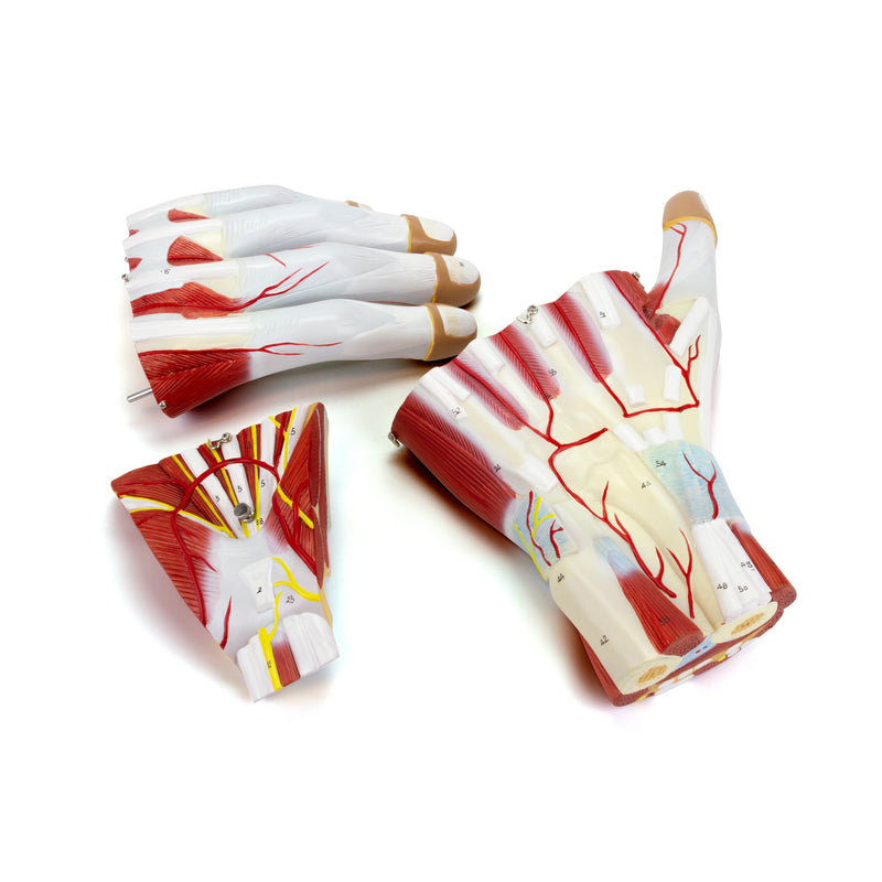
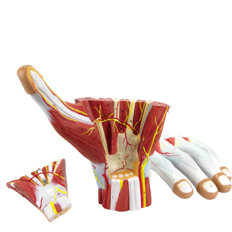
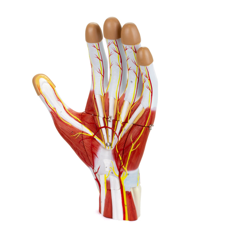
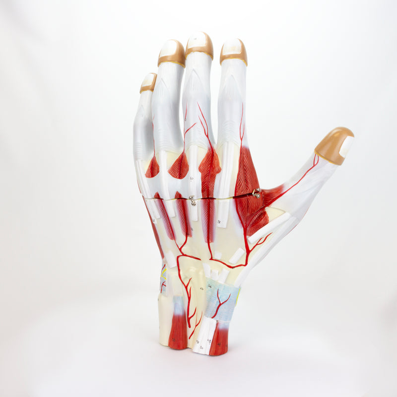
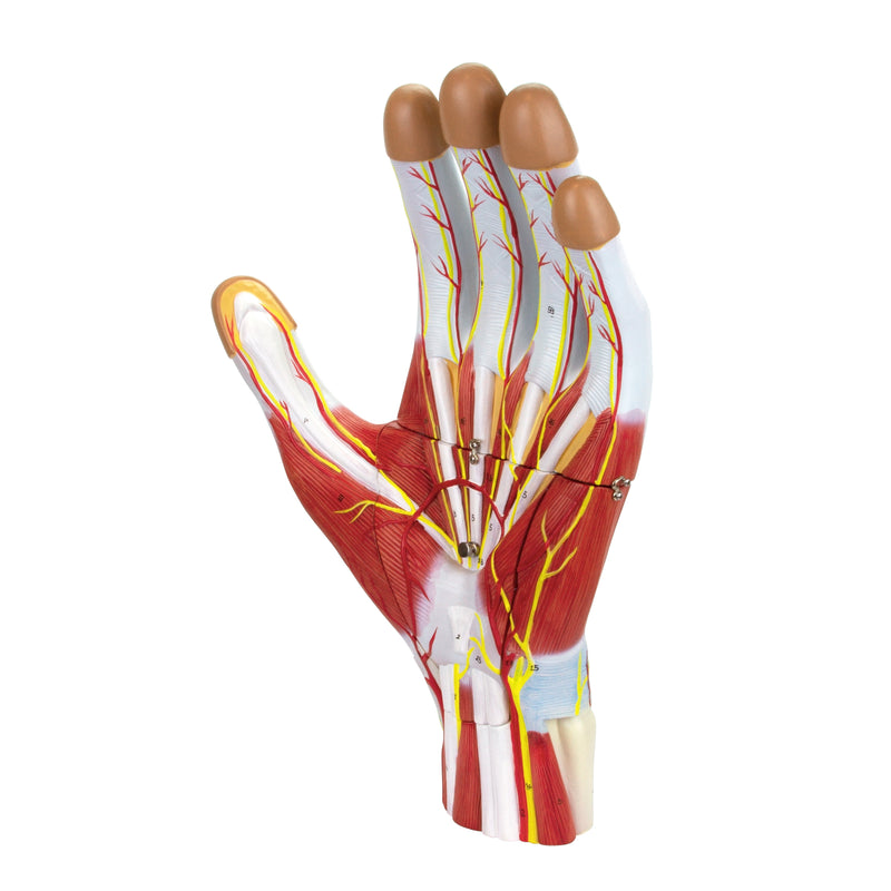
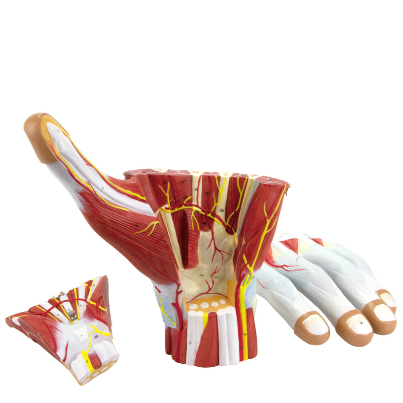
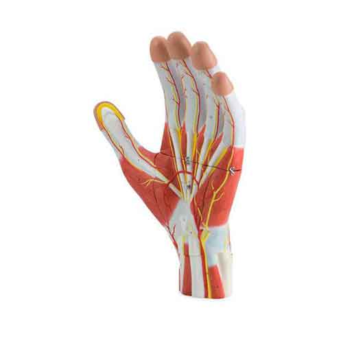
2배 확대된 이 모형으로 손의 해부학적 구조를 살펴보세요. 근육, 신경, 인대, 혈관 및 뼈 구조가 자세히 표현되어 있습니다. 제거 가능한 건막을 통해 내부 층을 심층적으로 학습할 수 있어 의료 교육 및 훈련에 적합합니다.
정보 카드로 활성화되는 증강 현실 기능을 통해 구조를 대화형 3D로 시각화하며 학습 경험을 향상할 수 있습니다. 피부색을 맞춤 설정할 수 있어 더욱 사실적이고 포괄적인 표현이 가능합니다.
대화형 3D 및 증강 현실 학습
제공되는 경우 함께 제공된 대화형 3D 및 증강 현실 자료를 사용하여 해부학적 구조를 더욱 자세히 관찰하고 비교해 보세요.
Hand Structure 2 Times Enlarged, 3 Parts Key Features
- Enlarged model (2x natural size) for better visualization of structures.
- Removable palmar aponeurosis for detailed study of internal layers.
- Detailed representation of anatomical structures.
Main Structures
- 1. Flexor carpi radialis muscle
- 2. Palmaris longus tendon
- 3. Flexor carpi ulnaris muscle
- 4. Flexor pollicis longus tendon
- 5. Flexor digitorum superficialis tendon
- 6. Flexor digitorum profundus tendon
- 7. Pronator quadratus muscle
- 8. Radial artery
- 9. Superficial palmar branch of radial artery
- 10. Dorsal carpal branch of radial artery
설명
손 구조, 2배 확대, 3개 부위
2배 확대된 이 모형으로 손의 해부학적 구조를 살펴보세요. 근육, 신경, 인대, 혈관 및 뼈 구조가 자세히 표현되어 있습니다. 제거 가능한 건막을 통해 내부 층을 심층적으로 학습할 수 있어 의료 교육 및 훈련에 적합합니다.
정보 카드로 활성화되는 증강 현실 기능을 통해 구조를 대화형 3D로 시각화하며 학습 경험을 향상할 수 있습니다. 피부색을 맞춤 설정할 수 있어 더욱 사실적이고 포괄적인 표현이 가능합니다.
대화형 3D 및 증강 현실 학습
제공되는 경우 함께 제공된 대화형 3D 및 증강 현실 자료를 사용하여 해부학적 구조를 더욱 자세히 관찰하고 비교해 보세요.
주요 특징
Hand Structure 2 Times Enlarged, 3 Parts Key Features
- Enlarged model (2x natural size) for better visualization of structures.
- Removable palmar aponeurosis for detailed study of internal layers.
- Detailed representation of anatomical structures.
Main Structures
- 1. Flexor carpi radialis muscle
- 2. Palmaris longus tendon
- 3. Flexor carpi ulnaris muscle
- 4. Flexor pollicis longus tendon
- 5. Flexor digitorum superficialis tendon
- 6. Flexor digitorum profundus tendon
- 7. Pronator quadratus muscle
- 8. Radial artery
- 9. Superficial palmar branch of radial artery
- 10. Dorsal carpal branch of radial artery
추가 정보
Invalid password
Enter
슬라이드쇼를 탐색하려면 왼쪽/오른쪽 화살표를 사용하거나 모바일 기기를 사용하는 경우 왼쪽/오른쪽으로 밀어 넘기세요