


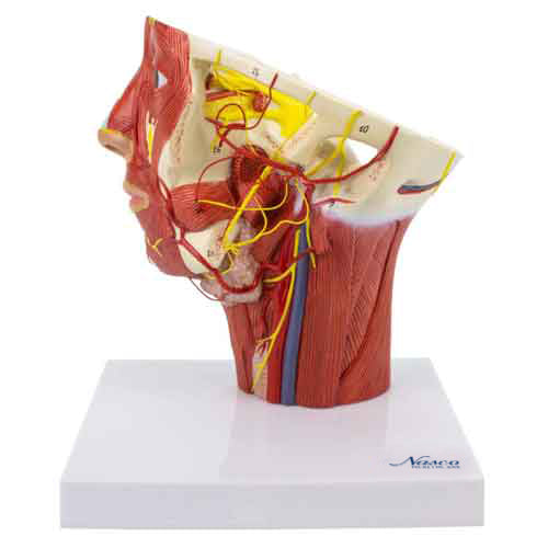
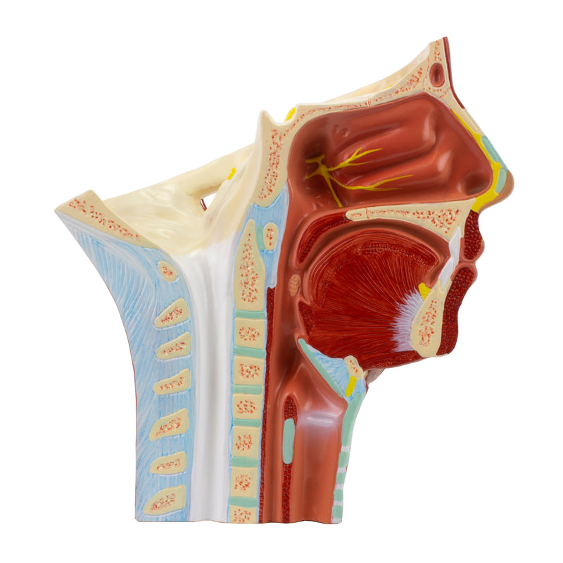
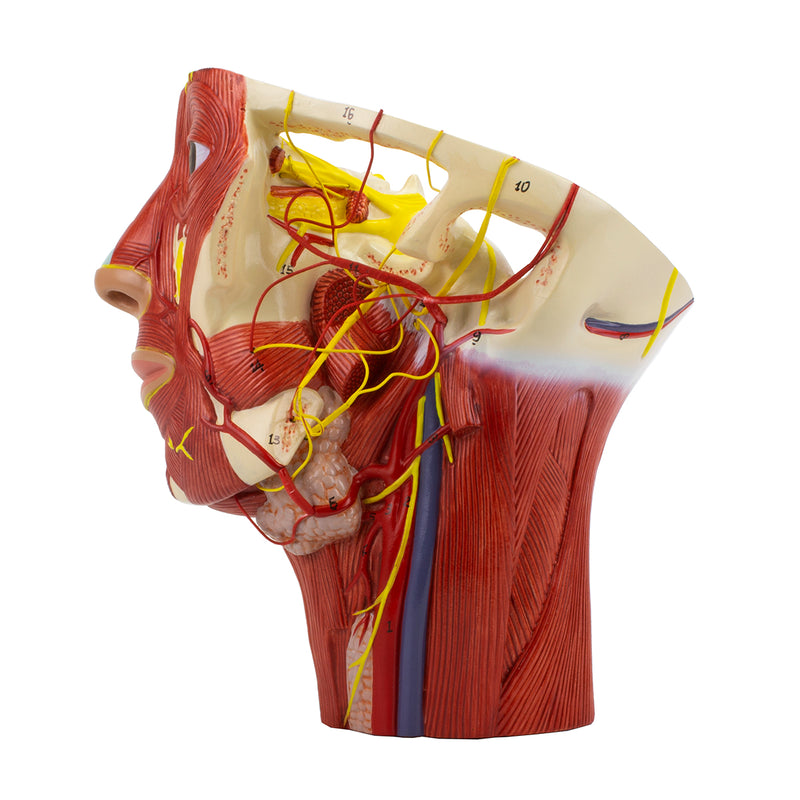
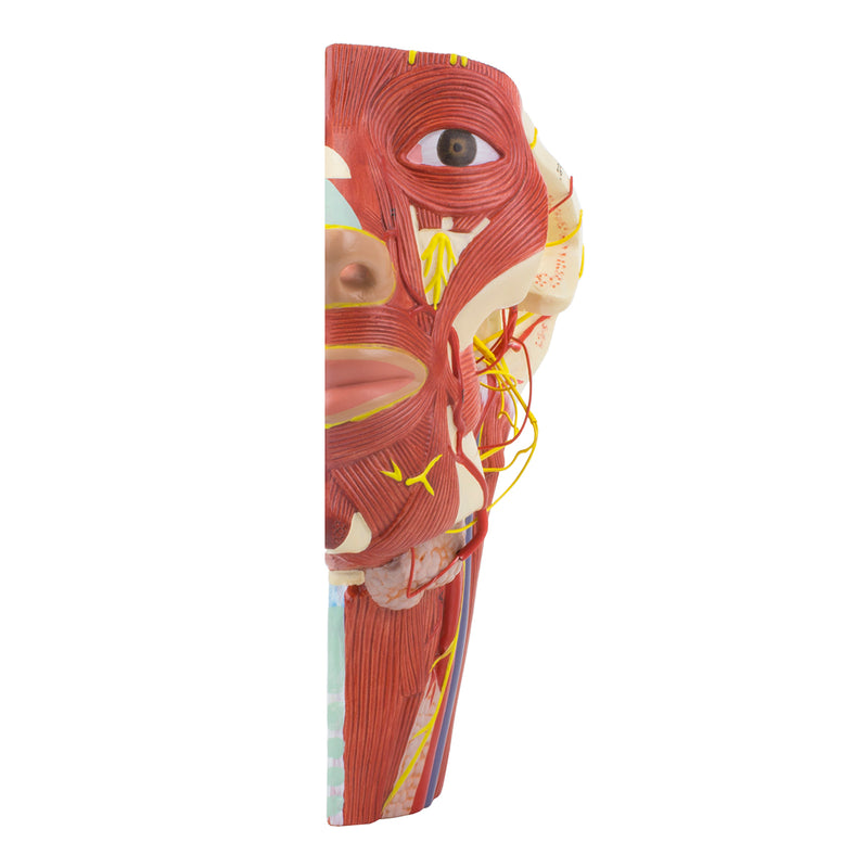
動脈付き頭部筋模型
左外頸動脈とその分枝を再現した等身大の解剖模型で、頭部と頸部の複雑な血管構造を詳しく学べます。拡張現実技術により、インタラクティブな3D表示で詳細かつ比較しながら学習でき、学生や医療従事者に最適です。
インタラクティブ3D&拡張現実学習
付属のインタラクティブ3Dおよび拡張現実リソースが提供されている場合は、それらを使用して解剖学的構造をより詳しく観察し、比較できます。
Head Muscles with Arteries Key Features
- Scale: Life-size
- Material: Resin
- Normal
- Tessellated Fundus
- Large Optic Disc Cupping
- DR1 (Diabetic Retinopathy - Stage 1)
- DR2 (Diabetic Retinopathy - Stage 2)
- DR3 (Diabetic Retinopathy - Stage 3)
Main Structures
- Common carotid artery
- Internal carotid artery
- External carotid artery
- Superior thyroid artery
- Lingual artery
- Facial artery
- Occipital artery
説明
動脈付き頭部筋模型
左外頸動脈とその分枝を再現した等身大の解剖模型で、頭部と頸部の複雑な血管構造を詳しく学べます。拡張現実技術により、インタラクティブな3D表示で詳細かつ比較しながら学習でき、学生や医療従事者に最適です。
インタラクティブ3D&拡張現実学習
付属のインタラクティブ3Dおよび拡張現実リソースが提供されている場合は、それらを使用して解剖学的構造をより詳しく観察し、比較できます。
主な特徴
Head Muscles with Arteries Key Features
- Scale: Life-size
- Material: Resin
- Normal
- Tessellated Fundus
- Large Optic Disc Cupping
- DR1 (Diabetic Retinopathy - Stage 1)
- DR2 (Diabetic Retinopathy - Stage 2)
- DR3 (Diabetic Retinopathy - Stage 3)
Main Structures
- Common carotid artery
- Internal carotid artery
- External carotid artery
- Superior thyroid artery
- Lingual artery
- Facial artery
- Occipital artery
追加情報
Invalid password
Enter
スライドショーを操作するには、左右の矢印を使うか、携帯端末をご利用の場合は左右にスワイプしてください