


視床下部付き脳幹、2倍拡大・4分割
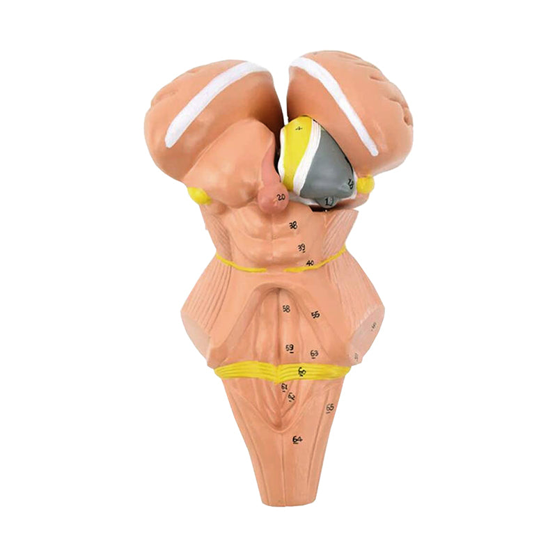
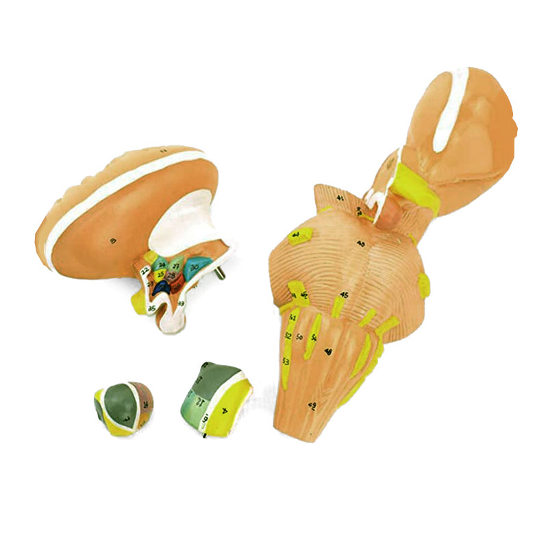
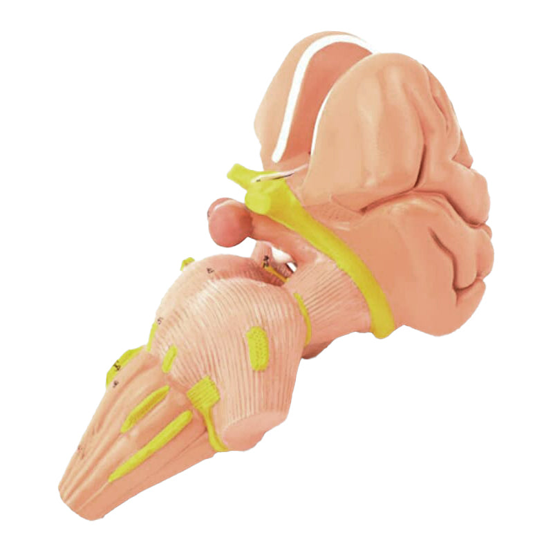
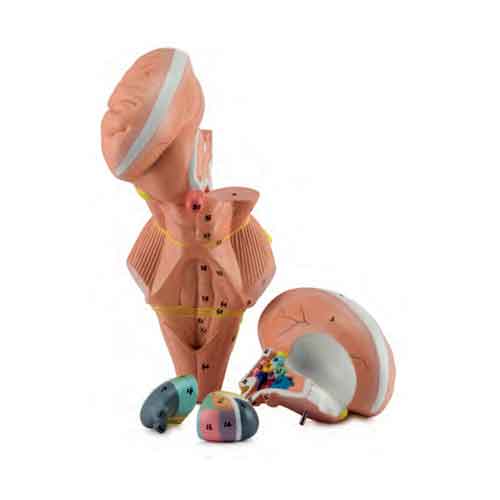
脳幹と視床下部核を実物の2倍に拡大した解剖模型で、4つのパーツに分かれているため、内部構造を詳しく観察できます。取り外し可能な視床と脳幹のパーツにより、視床下部核を明瞭に確認できます。
番号と色で示された脳神経および構造、情報カードが模型を補完します。情報カードを通じて拡張現実を体験でき、インタラクティブな3Dビジュアライゼーションにより、解剖学の学習と理解を深められます。
インタラクティブ3D&拡張現実学習
提供されている場合は、付属のインタラクティブ3Dおよび拡張現実リソースを使用して、解剖学的構造をより詳しく観察・比較できます。
Brain Sten with Hypothalamus, 2 Times Enlarged 4 Parts Key Features
- Anatomy study in schools and universities.
- Professional training.
- Patient explanations.
- Medical and scientific information.
- Neuroanatomy studies.
- Surgical dissection training.
- Patient education and procedure demonstration.
- Created from detailed prototypes.
Main Structures
- insula
- caudate nucleus
- internal capsule
- thalamus
- optic nerve (II)
- optic tract
- lateral geniculate body
- medial geniculate body
- pineal body
- superior colliculus
Product Specifications
- Scale: 2x life size
- Material: Synthetic resin
- Number of Parts: 4
- Assembly Type: No support base
説明
視床下部付き脳幹、2倍拡大・4分割
脳幹と視床下部核を実物の2倍に拡大した解剖模型で、4つのパーツに分かれているため、内部構造を詳しく観察できます。取り外し可能な視床と脳幹のパーツにより、視床下部核を明瞭に確認できます。
番号と色で示された脳神経および構造、情報カードが模型を補完します。情報カードを通じて拡張現実を体験でき、インタラクティブな3Dビジュアライゼーションにより、解剖学の学習と理解を深められます。
インタラクティブ3D&拡張現実学習
提供されている場合は、付属のインタラクティブ3Dおよび拡張現実リソースを使用して、解剖学的構造をより詳しく観察・比較できます。
主な特徴
Brain Sten with Hypothalamus, 2 Times Enlarged 4 Parts Key Features
- Anatomy study in schools and universities.
- Professional training.
- Patient explanations.
- Medical and scientific information.
- Neuroanatomy studies.
- Surgical dissection training.
- Patient education and procedure demonstration.
- Created from detailed prototypes.
Main Structures
- insula
- caudate nucleus
- internal capsule
- thalamus
- optic nerve (II)
- optic tract
- lateral geniculate body
- medial geniculate body
- pineal body
- superior colliculus
Product Specifications
- Scale: 2x life size
- Material: Synthetic resin
- Number of Parts: 4
- Assembly Type: No support base
追加情報
Invalid password
Enter
スライドショーを操作するには、左右の矢印を使うか、携帯端末をご利用の場合は左右にスワイプしてください