


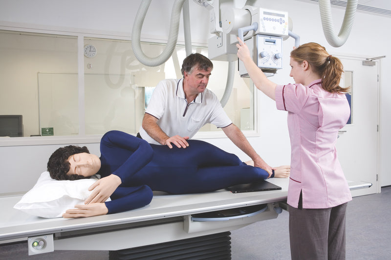
Il manichino radiografico per il posizionamento radiografico è un simulatore didattico a grandezza naturale progettato per insegnare il posizionamento radiografico, la gestione del paziente e le procedure di imaging senza esporre pazienti o studenti alle radiazioni. Ampiamente utilizzato nei programmi di radiografia, questo simulatore consente agli studenti di esercitarsi nelle corrette tecniche di posizionamento in un ambiente sicuro e controllato.
Il manichino contiene uno scheletro completamente articolato realizzato in plastica resistente e privo di componenti metallici, che ne consente l'utilizzo durante gli esami radiografici e le scansioni TC. Le strutture interne radiotrasparenti e l'anatomia superficiale realistica facilitano l'identificazione dei punti di riferimento anatomici durante la formazione sul posizionamento e sull'imaging.
Caratteristiche principali
• Manichino radiografico a grandezza naturale per il posizionamento
• Scheletro completamente flessibile e articolato
• Nessuna parte metallica, per un utilizzo sicuro con radiografie e TC
• Organi interni e strutture anatomiche radiotrasparenti
• Flessibilità articolare realistica per esercitarsi nel posizionamento
• Struttura resistente per un utilizzo didattico ripetuto
• Leggero e facile da maneggiare
Applicazioni didattiche
• Tecniche di posizionamento radiografico
• Formazione sulla gestione del paziente
• Posizionamento sulla macchina radiografica
• Esercitazioni sul posizionamento per TC
• Istruzione nei laboratori di imaging
• Programmi didattici di radiografia
Dettagli aggiuntivi
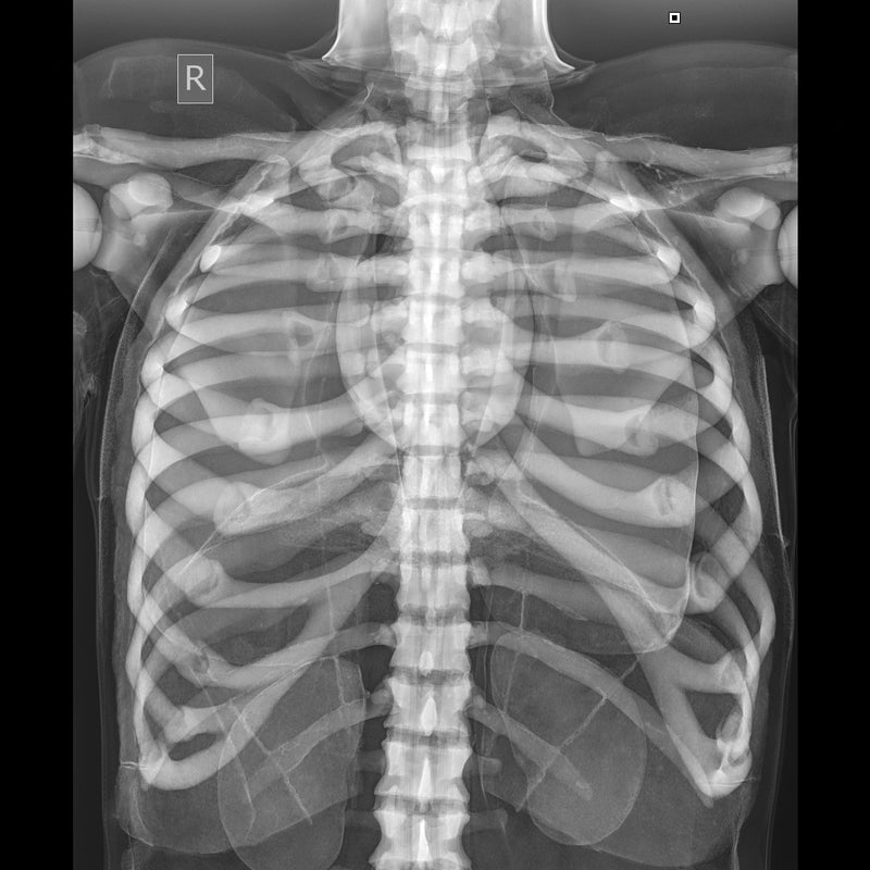
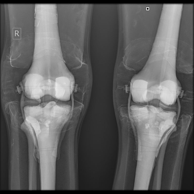
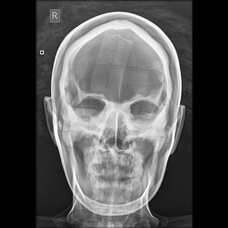
• Fornito con immagini radiografiche individuali del manichino
• Adatto per esercitarsi nel posizionamento del corpo intero
• Progettato per un utilizzo ripetuto in aula
Garanzia di 2 anni.
Peso di spedizione: 38 lbs
Dimensioni (pollici): 68.5 x 20.5 x 14
Key Features
• Life-size radiographic positioning manikin
• Fully flexible articulated skeleton
• No metal parts for safe use with X-ray and CT
• Radiolucent internal organs and anatomical structures
• Realistic joint flexibility for positioning practice
• Durable construction for repeated training use
• Lightweight for easy handling
Downloads
Individual X-Rays
Individual X-Rays for each Doll
- Each Doll is individually X-rayed
- Customers can be reassured that there are no metal parts in the Doll by referring to the set of digital X-
- Rays which are supplied with each Doll
- Images shown were taken using medium frequency generator; no grid; fine focus; 100cm S.I.D. Fuji CR system
Positioning and CT Scanning
Positioning
- The Doll is best used in the recumbent positions as, although it is possible to use an upright bucky with supports, this makes the use of positioning blocks and immobilization devices more difficult
- The Doll will lie naturally in the neutral position, prone or supine, without support
- A wide selection of positioning aids must be at hand, e.g. foam blocks, wedges, sand bags and compression band. These are essential to maintain any position other than neutral as the skin of the Doll has some resistance to overcome. This is, of course, an advantage in the teaching as it prevents the student cutting corners by omitting the use of these positioning aids
- The Doll can be positioned for all standard projections with no more difficulty than a difficult patient might present.
CT Scanning
- The X-Ray Positioning Doll was positioned and imaged for a whole body CT (Computed Tomography) investigation
- Good images in terms of body outline and fundamental structure can be obtained
- Certain areas of the skeleton structure e.g. thoracic vertebrae and the head of the femur gave similar values to the typical patient
- As the doll is very different from the human body in terms of attenuation, it cannot be used for training in, or practising exposure control
Il manichino radiografico per il posizionamento radiografico è un simulatore didattico a grandezza naturale progettato per insegnare il posizionamento radiografico, la gestione del paziente e le procedure di imaging senza esporre pazienti o studenti alle radiazioni. Ampiamente utilizzato nei programmi di radiografia, questo simulatore consente agli studenti di esercitarsi nelle corrette tecniche di posizionamento in un ambiente sicuro e controllato.
Il manichino contiene uno scheletro completamente articolato realizzato in plastica resistente e privo di componenti metallici, che ne consente l'utilizzo durante gli esami radiografici e le scansioni TC. Le strutture interne radiotrasparenti e l'anatomia superficiale realistica facilitano l'identificazione dei punti di riferimento anatomici durante la formazione sul posizionamento e sull'imaging.
Caratteristiche principali
• Manichino radiografico a grandezza naturale per il posizionamento
• Scheletro completamente flessibile e articolato
• Nessuna parte metallica, per un utilizzo sicuro con radiografie e TC
• Organi interni e strutture anatomiche radiotrasparenti
• Flessibilità articolare realistica per esercitarsi nel posizionamento
• Struttura resistente per un utilizzo didattico ripetuto
• Leggero e facile da maneggiare
Applicazioni didattiche
• Tecniche di posizionamento radiografico
• Formazione sulla gestione del paziente
• Posizionamento sulla macchina radiografica
• Esercitazioni sul posizionamento per TC
• Istruzione nei laboratori di imaging
• Programmi didattici di radiografia
Dettagli aggiuntivi
• Fornito con immagini radiografiche individuali del manichino
• Adatto per esercitarsi nel posizionamento del corpo intero
• Progettato per un utilizzo ripetuto in aula
Garanzia di 2 anni.
Peso di spedizione: 38 lbs
Dimensioni (pollici): 68.5 x 20.5 x 14
Key Features
• Life-size radiographic positioning manikin
• Fully flexible articulated skeleton
• No metal parts for safe use with X-ray and CT
• Radiolucent internal organs and anatomical structures
• Realistic joint flexibility for positioning practice
• Durable construction for repeated training use
• Lightweight for easy handling
Downloads
Individual X-Rays
Individual X-Rays for each Doll
- Each Doll is individually X-rayed
- Customers can be reassured that there are no metal parts in the Doll by referring to the set of digital X-
- Rays which are supplied with each Doll
- Images shown were taken using medium frequency generator; no grid; fine focus; 100cm S.I.D. Fuji CR system
Positioning and CT Scanning
Positioning
- The Doll is best used in the recumbent positions as, although it is possible to use an upright bucky with supports, this makes the use of positioning blocks and immobilization devices more difficult
- The Doll will lie naturally in the neutral position, prone or supine, without support
- A wide selection of positioning aids must be at hand, e.g. foam blocks, wedges, sand bags and compression band. These are essential to maintain any position other than neutral as the skin of the Doll has some resistance to overcome. This is, of course, an advantage in the teaching as it prevents the student cutting corners by omitting the use of these positioning aids
- The Doll can be positioned for all standard projections with no more difficulty than a difficult patient might present.
CT Scanning
- The X-Ray Positioning Doll was positioned and imaged for a whole body CT (Computed Tomography) investigation
- Good images in terms of body outline and fundamental structure can be obtained
- Certain areas of the skeleton structure e.g. thoracic vertebrae and the head of the femur gave similar values to the typical patient
- As the doll is very different from the human body in terms of attenuation, it cannot be used for training in, or practising exposure control