


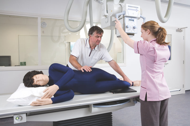
Le mannequin de positionnement radiographique pour rayons X est un simulateur d’entraînement grandeur nature conçu pour l’enseignement du positionnement radiographique, de la manipulation des patients et des procédures d’imagerie, sans exposer les patients ni les étudiants aux rayonnements. Largement utilisé par les programmes de radiographie, ce simulateur permet aux apprenants de s’exercer aux techniques de positionnement appropriées dans un environnement sûr et contrôlé.
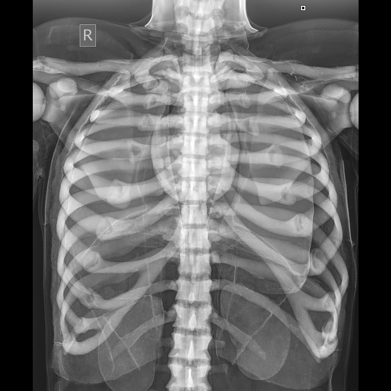
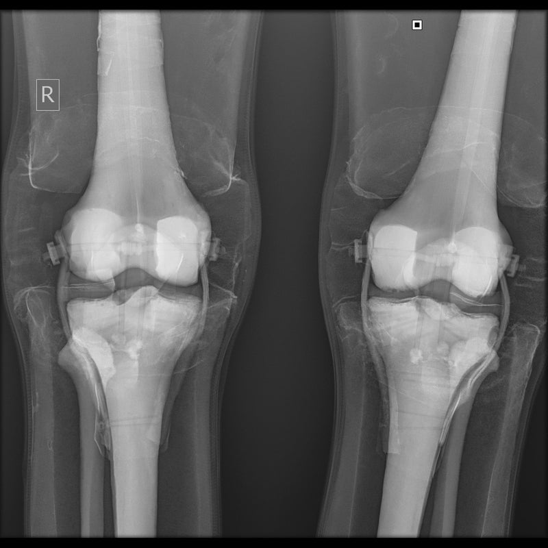
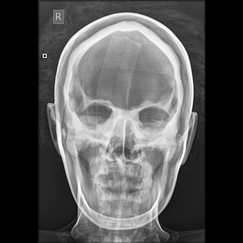
Le mannequin contient un squelette entièrement articulé fabriqué en plastique durable, sans composants métalliques, ce qui permet de l’utiliser lors d’examens aux rayons X et de tomodensitométrie. Les structures internes radiotransparentes et l’anatomie de surface réaliste facilitent l’identification des repères anatomiques pendant la formation au positionnement et à l’imagerie.
Caractéristiques principales
• Mannequin de positionnement radiographique grandeur nature
• Squelette articulé entièrement flexible
• Aucune pièce métallique pour une utilisation sûre avec les rayons X et la tomodensitométrie
• Organes et structures anatomiques internes radiotransparents
• Flexibilité articulaire réaliste pour la pratique du positionnement
• Construction durable pour des utilisations répétées en formation
• Léger et facile à manipuler
Applications de formation
• Techniques de positionnement radiographique
• Formation à la manipulation des patients
• Positionnement sur les appareils à rayons X
• Pratique du positionnement en tomodensitométrie
• Enseignement en laboratoire d’imagerie
• Programmes de formation en radiographie
Informations complémentaires
• Fourni avec des images radiographiques individuelles du mannequin
• Convient à la pratique du positionnement du corps entier
• Conçu pour une utilisation répétée en classe
Garantie de 2 ans.
Poids d’expédition : 38 lbs
Dimensions (pouces) : 68.5 x 20.5 x 14
Key Features
• Life-size radiographic positioning manikin
• Fully flexible articulated skeleton
• No metal parts for safe use with X-ray and CT
• Radiolucent internal organs and anatomical structures
• Realistic joint flexibility for positioning practice
• Durable construction for repeated training use
• Lightweight for easy handling
Downloads
Individual X-Rays
Individual X-Rays for each Doll
- Each Doll is individually X-rayed
- Customers can be reassured that there are no metal parts in the Doll by referring to the set of digital X-
- Rays which are supplied with each Doll
- Images shown were taken using medium frequency generator; no grid; fine focus; 100cm S.I.D. Fuji CR system
Positioning and CT Scanning
Positioning
- The Doll is best used in the recumbent positions as, although it is possible to use an upright bucky with supports, this makes the use of positioning blocks and immobilization devices more difficult
- The Doll will lie naturally in the neutral position, prone or supine, without support
- A wide selection of positioning aids must be at hand, e.g. foam blocks, wedges, sand bags and compression band. These are essential to maintain any position other than neutral as the skin of the Doll has some resistance to overcome. This is, of course, an advantage in the teaching as it prevents the student cutting corners by omitting the use of these positioning aids
- The Doll can be positioned for all standard projections with no more difficulty than a difficult patient might present.
CT Scanning
- The X-Ray Positioning Doll was positioned and imaged for a whole body CT (Computed Tomography) investigation
- Good images in terms of body outline and fundamental structure can be obtained
- Certain areas of the skeleton structure e.g. thoracic vertebrae and the head of the femur gave similar values to the typical patient
- As the doll is very different from the human body in terms of attenuation, it cannot be used for training in, or practising exposure control
Le mannequin de positionnement radiographique pour rayons X est un simulateur d’entraînement grandeur nature conçu pour l’enseignement du positionnement radiographique, de la manipulation des patients et des procédures d’imagerie, sans exposer les patients ni les étudiants aux rayonnements. Largement utilisé par les programmes de radiographie, ce simulateur permet aux apprenants de s’exercer aux techniques de positionnement appropriées dans un environnement sûr et contrôlé.
Le mannequin contient un squelette entièrement articulé fabriqué en plastique durable, sans composants métalliques, ce qui permet de l’utiliser lors d’examens aux rayons X et de tomodensitométrie. Les structures internes radiotransparentes et l’anatomie de surface réaliste facilitent l’identification des repères anatomiques pendant la formation au positionnement et à l’imagerie.
Caractéristiques principales
• Mannequin de positionnement radiographique grandeur nature
• Squelette articulé entièrement flexible
• Aucune pièce métallique pour une utilisation sûre avec les rayons X et la tomodensitométrie
• Organes et structures anatomiques internes radiotransparents
• Flexibilité articulaire réaliste pour la pratique du positionnement
• Construction durable pour des utilisations répétées en formation
• Léger et facile à manipuler
Applications de formation
• Techniques de positionnement radiographique
• Formation à la manipulation des patients
• Positionnement sur les appareils à rayons X
• Pratique du positionnement en tomodensitométrie
• Enseignement en laboratoire d’imagerie
• Programmes de formation en radiographie
Informations complémentaires
• Fourni avec des images radiographiques individuelles du mannequin
• Convient à la pratique du positionnement du corps entier
• Conçu pour une utilisation répétée en classe
Garantie de 2 ans.
Poids d’expédition : 38 lbs
Dimensions (pouces) : 68.5 x 20.5 x 14
Key Features
• Life-size radiographic positioning manikin
• Fully flexible articulated skeleton
• No metal parts for safe use with X-ray and CT
• Radiolucent internal organs and anatomical structures
• Realistic joint flexibility for positioning practice
• Durable construction for repeated training use
• Lightweight for easy handling
Downloads
Individual X-Rays
Individual X-Rays for each Doll
- Each Doll is individually X-rayed
- Customers can be reassured that there are no metal parts in the Doll by referring to the set of digital X-
- Rays which are supplied with each Doll
- Images shown were taken using medium frequency generator; no grid; fine focus; 100cm S.I.D. Fuji CR system
Positioning and CT Scanning
Positioning
- The Doll is best used in the recumbent positions as, although it is possible to use an upright bucky with supports, this makes the use of positioning blocks and immobilization devices more difficult
- The Doll will lie naturally in the neutral position, prone or supine, without support
- A wide selection of positioning aids must be at hand, e.g. foam blocks, wedges, sand bags and compression band. These are essential to maintain any position other than neutral as the skin of the Doll has some resistance to overcome. This is, of course, an advantage in the teaching as it prevents the student cutting corners by omitting the use of these positioning aids
- The Doll can be positioned for all standard projections with no more difficulty than a difficult patient might present.
CT Scanning
- The X-Ray Positioning Doll was positioned and imaged for a whole body CT (Computed Tomography) investigation
- Good images in terms of body outline and fundamental structure can be obtained
- Certain areas of the skeleton structure e.g. thoracic vertebrae and the head of the femur gave similar values to the typical patient
- As the doll is very different from the human body in terms of attenuation, it cannot be used for training in, or practising exposure control