


Befruchtung und embryonale Entwicklung
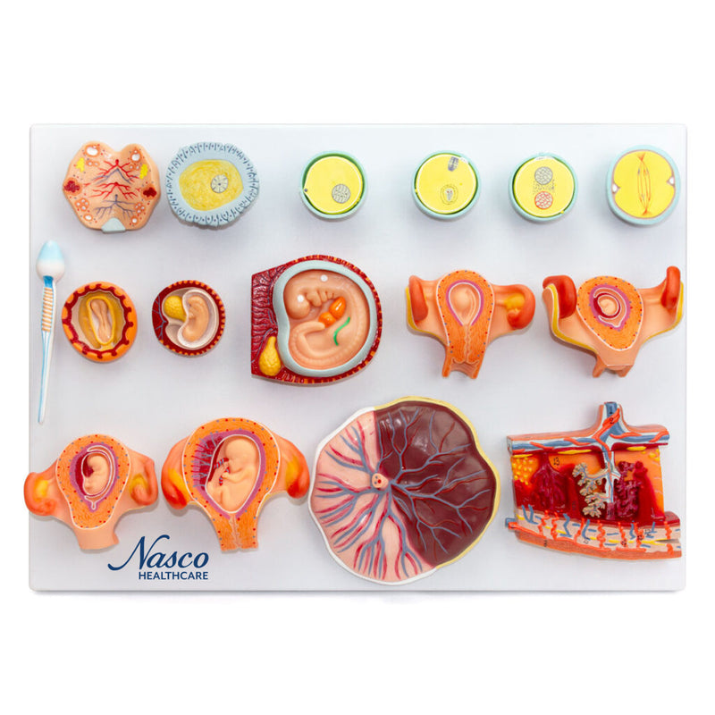
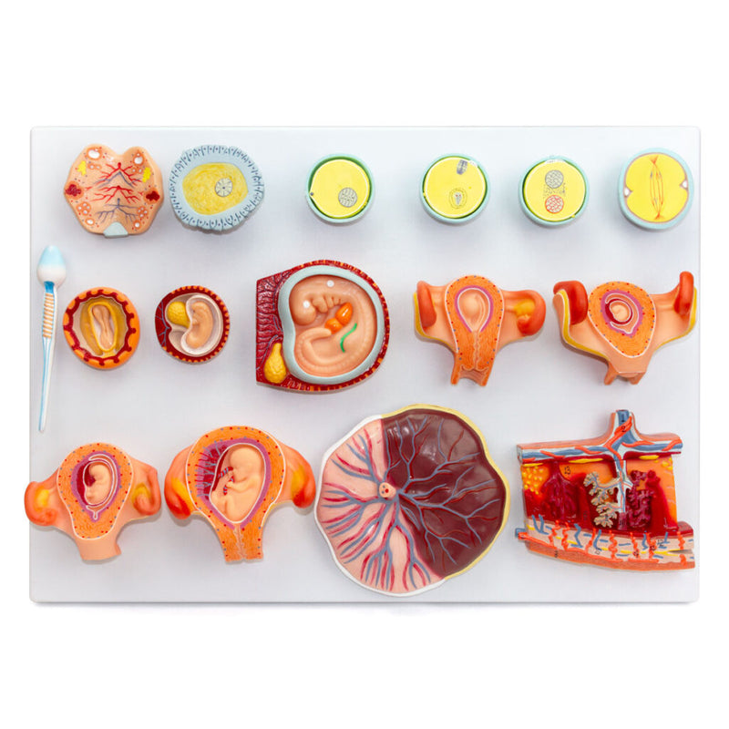
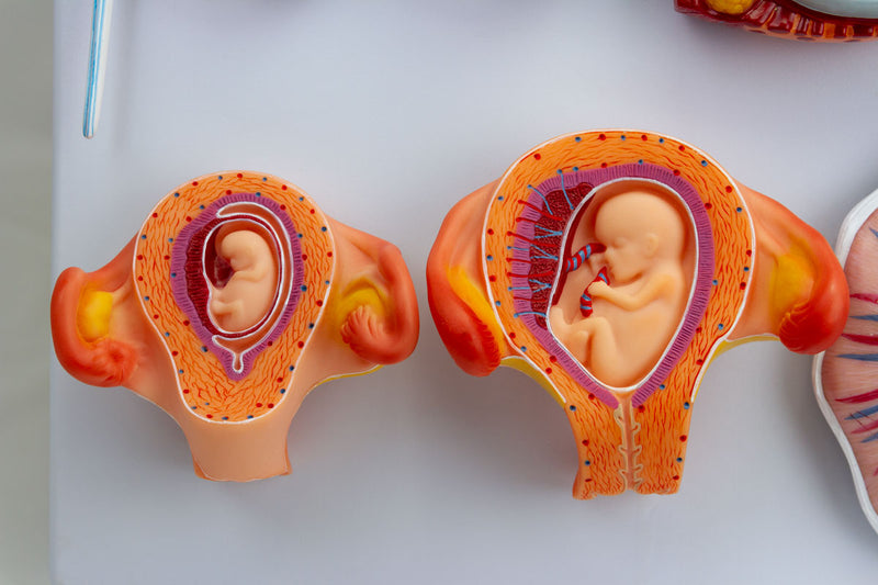
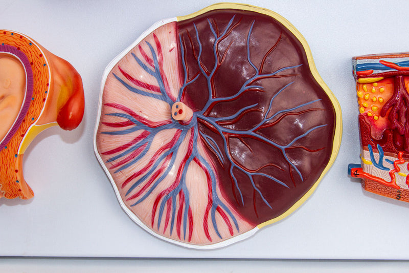
Dieses detaillierte anatomische Modell zeigt 16 Stadien der menschlichen Befruchtung und embryonalen Entwicklung – von der Vereinigung der Gameten bis zum Fötus im dritten Monat, einschließlich der Plazenta. Das Modell besteht aus hochwertigem Kunstharz, ist handbemalt und präzise nummeriert, auf einer Polymerbasis montiert und enthält eine Informationskarte mit Augmented Reality (AR).
Die interaktive 3D-Visualisierung über AR ergänzt das Lernen und ermöglicht eine detaillierte Analyse von Strukturen wie Vorkernen, Zygote, Blastoderm und Embryo in verschiedenen Stadien. Ideal für Ausbildung und Forschung in medizinischen und biologischen Fachbereichen.
Interaktives 3D- und Augmented-Reality-Lernen
Nutzen Sie, sofern verfügbar, die begleitenden interaktiven 3D- und Augmented-Reality-Ressourcen, um anatomische Strukturen detaillierter zu untersuchen und zu vergleichen.
Fertilization and Embryonic Development Key Features
- Detailed representation of each stage of embryonic development.
- Allows the study of the structures that make up the placenta.
- High-precision natural molding.
- Made of stable and resistant synthetic material.
- Original, numbered, and hand-painted replicas.
- Resin approved in toxicological tests.
Main Structures
- Mitosis
- Female pronucleus close to male pronucleus
- Female pronucleus and male pronucleus
- Amphimixis or zygote
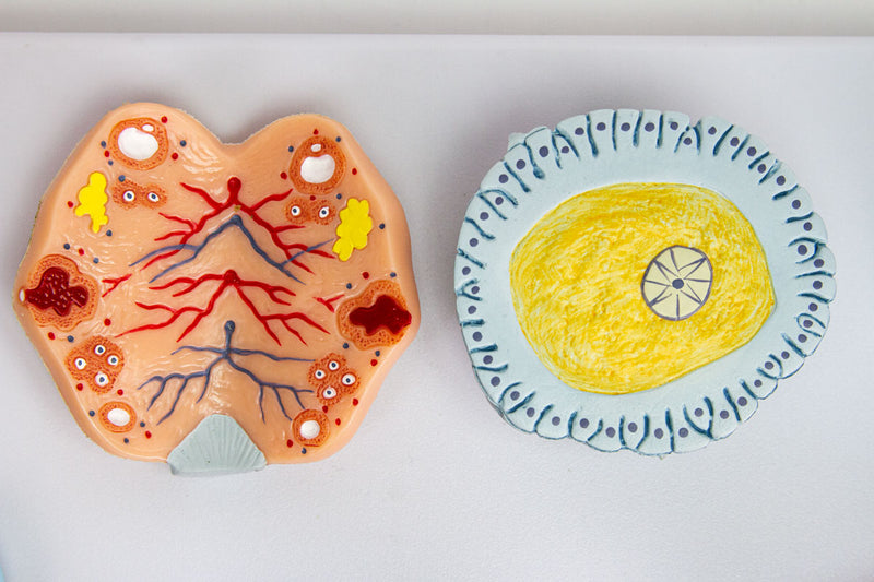
- Ovum
- Ovary
- Spermatozoon
- Blastoderm
- Embryo at week 2-3
- Embryo at week 5
Product Specifications
- Material: Synthetic resin.
- Scale: Unspecified.
- Manual numbering and painting.
- Information card included.
Befruchtung und embryonale Entwicklung
Dieses detaillierte anatomische Modell zeigt 16 Stadien der menschlichen Befruchtung und embryonalen Entwicklung – von der Vereinigung der Gameten bis zum Fötus im dritten Monat, einschließlich der Plazenta. Das Modell besteht aus hochwertigem Kunstharz, ist handbemalt und präzise nummeriert, auf einer Polymerbasis montiert und enthält eine Informationskarte mit Augmented Reality (AR).
Die interaktive 3D-Visualisierung über AR ergänzt das Lernen und ermöglicht eine detaillierte Analyse von Strukturen wie Vorkernen, Zygote, Blastoderm und Embryo in verschiedenen Stadien. Ideal für Ausbildung und Forschung in medizinischen und biologischen Fachbereichen.
Interaktives 3D- und Augmented-Reality-Lernen
Nutzen Sie, sofern verfügbar, die begleitenden interaktiven 3D- und Augmented-Reality-Ressourcen, um anatomische Strukturen detaillierter zu untersuchen und zu vergleichen.
Fertilization and Embryonic Development Key Features
- Detailed representation of each stage of embryonic development.
- Allows the study of the structures that make up the placenta.
- High-precision natural molding.
- Made of stable and resistant synthetic material.
- Original, numbered, and hand-painted replicas.
- Resin approved in toxicological tests.
Main Structures
- Mitosis
- Female pronucleus close to male pronucleus
- Female pronucleus and male pronucleus
- Amphimixis or zygote
- Ovum
- Ovary
- Spermatozoon
- Blastoderm
- Embryo at week 2-3
- Embryo at week 5
Product Specifications
- Material: Synthetic resin.
- Scale: Unspecified.
- Manual numbering and painting.
- Information card included.