


Fish Anatomy, 5 Parts
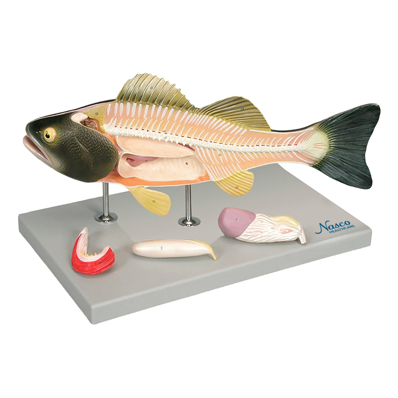
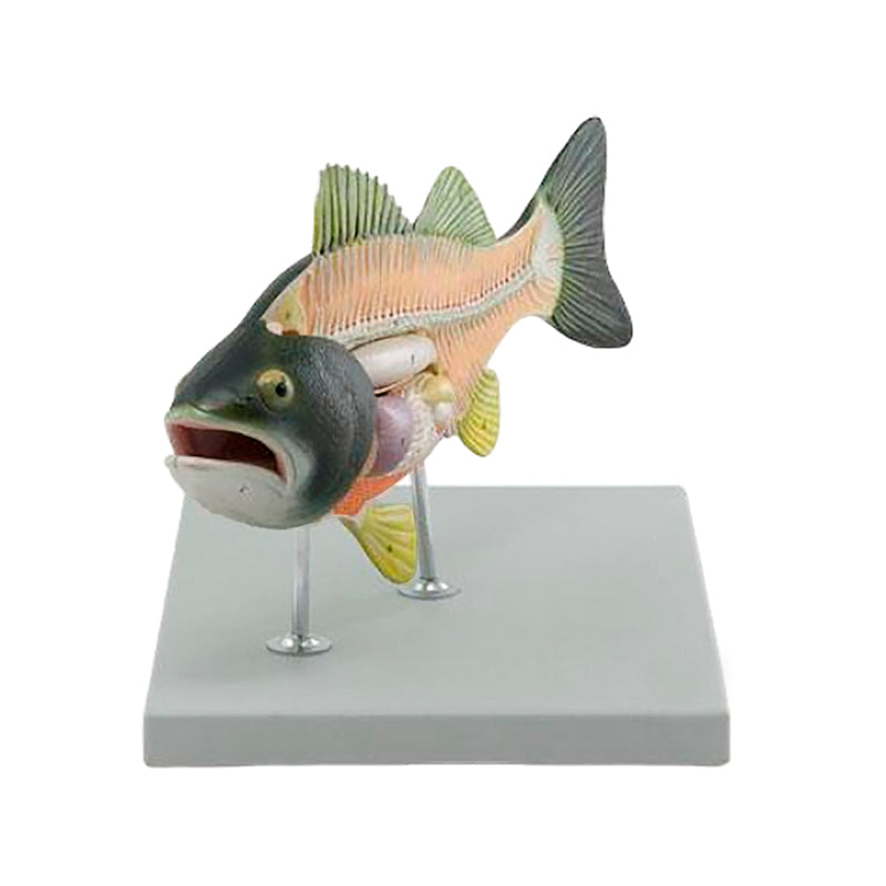
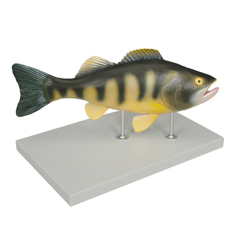
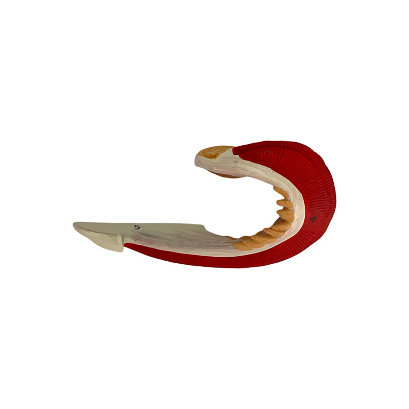
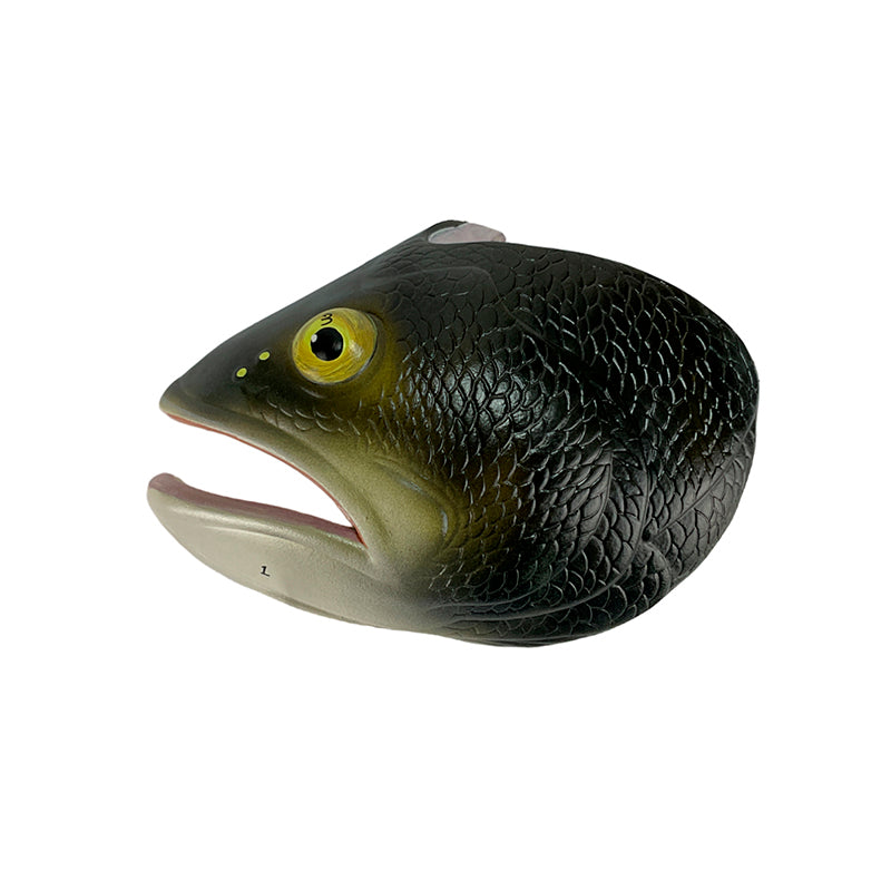
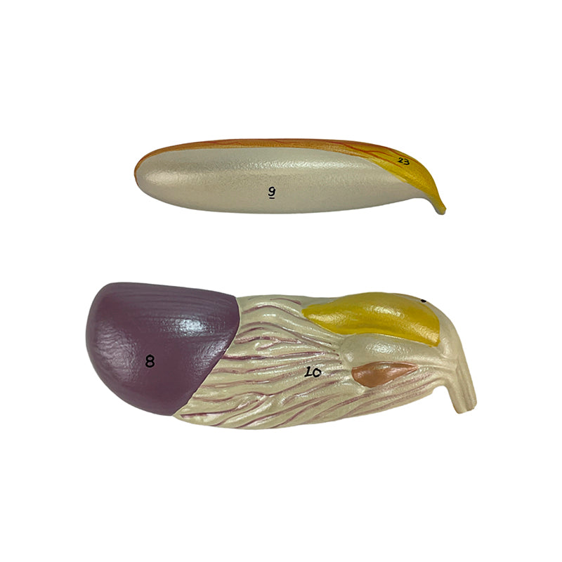
This dissected anatomical model of a perch (Perca sp.) allows for detailed study of the internal and external anatomy of a bony fish. With 5 removable parts secured by magnets, it offers individualized analysis of structures such as the head, gill arch, liver, intestine, and swim bladder.
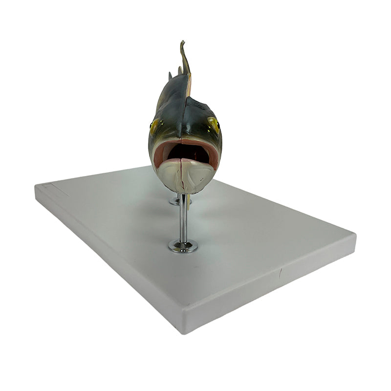
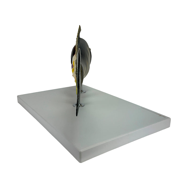
The right side shows the external anatomy, while the left reveals the internal anatomy through a longitudinal section. Made of high-precision synthetic resin, hand-painted and numbered, it includes a base, metal rods, and an information card with augmented reality (AR) for interactive 3D visualization, enhancing learning. Ideal for students and professionals in biology and veterinary science.
Interactive 3D & Augmented Reality Learning
Use the accompanying interactive 3D and augmented reality resources, where provided, to examine and compare anatomical structures in greater detail.
Fish Anatomy, 5 Parts Key Features
- Magnet clasps, facilitating assembly and disassembly;
- Metal rods and polymer base with screw and wing nut system for fixing;
- High precision molding;
- Made of stable synthetic material and resin, approved in toxicological tests;
- Detailed replicas;
- Numbered and hand-painted model;
- Includes polymer base, rods and metal support;
- Scale: (The scale needs to be extracted from the input text, if available. If not present, this
Main Structures
- ventral fin
- intestine
- liver
- anal fin
- hemal arch
- gas bladder
- spinous process
- centrum
- basapophysis
- cranium
Product Specifications
- Scale: (The scale needs to be extracted from the input text, if available. If not present, this
- Material: Synthetic resin.
Fish Anatomy, 5 Parts
This dissected anatomical model of a perch (Perca sp.) allows for detailed study of the internal and external anatomy of a bony fish. With 5 removable parts secured by magnets, it offers individualized analysis of structures such as the head, gill arch, liver, intestine, and swim bladder.
The right side shows the external anatomy, while the left reveals the internal anatomy through a longitudinal section. Made of high-precision synthetic resin, hand-painted and numbered, it includes a base, metal rods, and an information card with augmented reality (AR) for interactive 3D visualization, enhancing learning. Ideal for students and professionals in biology and veterinary science.
Interactive 3D & Augmented Reality Learning
Use the accompanying interactive 3D and augmented reality resources, where provided, to examine and compare anatomical structures in greater detail.
Fish Anatomy, 5 Parts Key Features
- Magnet clasps, facilitating assembly and disassembly;
- Metal rods and polymer base with screw and wing nut system for fixing;
- High precision molding;
- Made of stable synthetic material and resin, approved in toxicological tests;
- Detailed replicas;
- Numbered and hand-painted model;
- Includes polymer base, rods and metal support;
- Scale: (The scale needs to be extracted from the input text, if available. If not present, this
Main Structures
- ventral fin
- intestine
- liver
- anal fin
- hemal arch
- gas bladder
- spinous process
- centrum
- basapophysis
- cranium
Product Specifications
- Scale: (The scale needs to be extracted from the input text, if available. If not present, this
- Material: Synthetic resin.